Figures & data
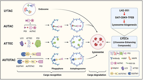
Figure 1. A schematic illustration of LYTAC, AUTAC, ATTEC, AUTOTAC and LYECs. LYTACs mark the extracellular POI with mannose-6-phosphate (M6P)-conjugated antibodies for lysosomal degradation. AUTACs contain a degradation tag (guanine derivative), which triggers K63 polyubiquitination of the POI, and the POI is recognized by the selective macroautophagy/autophagy pathway for degradation. ATTECs tether the POI to the phagophore for autophagic degradation by direct binding to the POI and LC3. AUTOTACs bring the POI to the phagophore for autophagic degradation by binding to the POI and SQSTM1/p62. LYECs promote activation of TFEB and lysosome biogenesis, which enhances the degradation efficiency of autolysosomes (created with BioRender.Com).