Figures & data
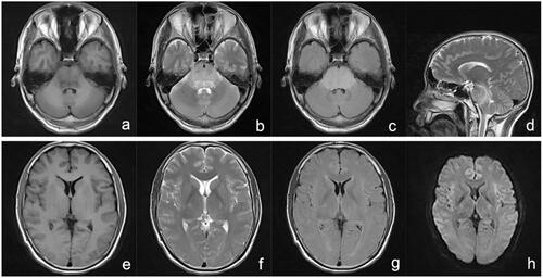
Figure 1. MRI 18 days after ingestion showing abnormal heterogeneous intensities on T1WI (a), T2WI (b and h), T2-FLAIR (c), and DWI (d) of the brainstem, particularly in the pons, in accordance with central pontine myelinolysis signal intensity. The bilateral caudate nucleus, putamen, and capsula externa showed abnormal low-intensity on T1WI (e), abnormal high-intensity on T2WI (f), and T2-FLAIR (g). DWI: diffusion-weighted imaging; FLAIR: fluid attenuated inversion recovery; MRI: magnetic resonance imaging.
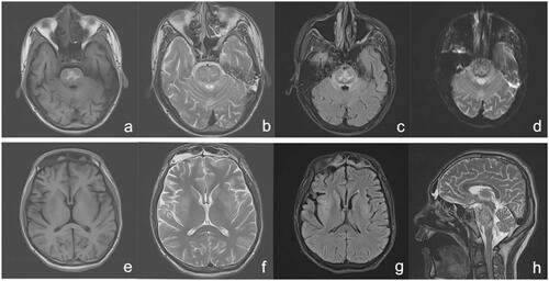
Figure 2. MRI 13 days after ingestion showing abnormal high-intensity in the pons, bilateral brachium pontis, and pedunculus cerebri on T1WI (a), T2WI (b and h), and T2-FLAIR (c), and slight high-intensity on DWI (d); The bilateral caudate nucleus, putamen, thalamus, and corona radiata showed abnormal low-intensity on T1WI (e) and abnormal high-intensity on T2WI (f) and T2-FLAIR (g). DWI: diffusion-weighted imaging; FLAIR: fluid attenuated inversion recovery; MRI: magnetic resonance imaging.
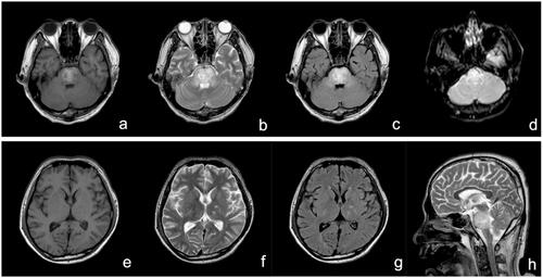
Figure 3. MRI 4 days after ingestion: T1WI (a and e), T2WI (b, d, and f), T2-FLAIR (c and g), and DWI (h) depicting an abnormal medulla oblongata, pons, midbrain, cerebellum, left thalamus, surrounding posterior horn of the lateral ventricle, bilateral brachium pontis, and pedunculus cerebri. DWI: diffusion-weighted imaging; FLAIR: fluid attenuated inversion recovery; MRI: magnetic resonance imaging.