1,515
Views
50
CrossRef citations to date
0
Altmetric
Case Report

Reversible myocardial injury associated with aluminum phosphide poisoning

, , , , , & show all
Pages 728-731 | Received 07 May 2006, Accepted 29 Nov 2006, Published online: 20 Jan 2009

Figures & data

Fig. 1. Electrocardiogram at admission of the first case showing ST-segment elevation in the leads V1 and V2.
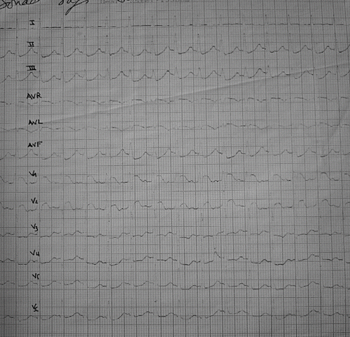
Fig. 1. Electrocardiogram at admission of the first case showing ST-segment elevation in the leads V1 and V2.

Fig. 2. Electrocardiogram of the first patient four hours after admission showed an increase of ST-segment in the leads V1 and V2.
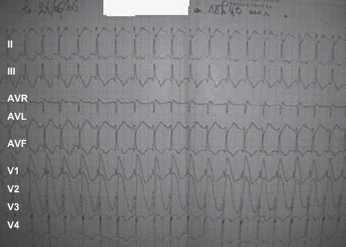
Fig. 2. Electrocardiogram of the first patient four hours after admission showed an increase of ST-segment in the leads V1 and V2.

Fig. 3. Electrocardiogram in admission of the second patient showing ST-segment elevated in leads V1, V2, and V3.
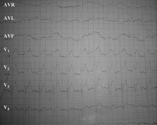
Fig. 3. Electrocardiogram in admission of the second patient showing ST-segment elevated in leads V1, V2, and V3.

Fig. 4. Echocardiographically parasternal long axis depicted improvement of ventricle function in the second case. A) Echocardiogram obtained on the second day after admission depicts global hypokinesis with LVEF of 30% (calculated by Simpson method) and dilatation of LV. B) Echocardiogram taken eight days after admission showed improvement of ventricle function LVEF of 30%. RV: right ventricle; IVS: interventricular septum; LV: left ventricle; PW: posterior wall.
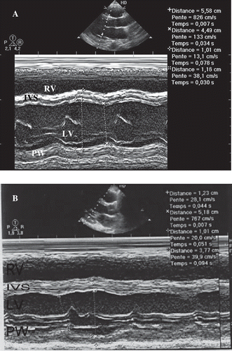
Fig. 4. Echocardiographically parasternal long axis depicted improvement of ventricle function in the second case. A) Echocardiogram obtained on the second day after admission depicts global hypokinesis with LVEF of 30% (calculated by Simpson method) and dilatation of LV. B) Echocardiogram taken eight days after admission showed improvement of ventricle function LVEF of 30%. RV: right ventricle; IVS: interventricular septum; LV: left ventricle; PW: posterior wall.

Reprints and Corporate Permissions

Please note: Selecting permissions does not provide access to the full text of the article, please see our help page How do I view content?

To request a reprint or corporate permissions for this article, please click on the relevant link below:

Academic Permissions

Please note: Selecting permissions does not provide access to the full text of the article, please see our help page How do I view content?

Obtain permissions instantly via Rightslink by clicking on the button below:

If you are unable to obtain permissions via Rightslink, please complete and submit this Permissions form. For more information, please visit our Permissions help page.