Figures & data
Fig. 1. MRI of the brain 12 hours after concentrated hydrogen peroxide ingestion demonstrating hyperintensities in (A) the right occipital cortex, left central sulcus and bilateral frontal lobes and in bilateral cerebellar hemispheres (B) on FLAIR imaging, areas of restricted diffusion in the bilateral cerebellar hemispheres (C) and in the right occipital cortex (D).
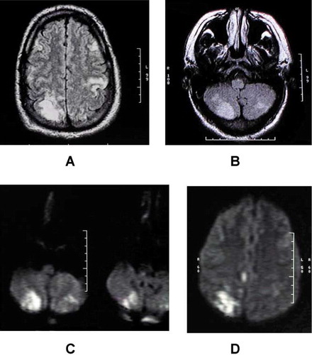
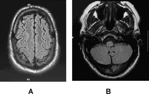
Fig. 2. MRI of the brain obtained 6 months after treatment with hyperbaric oxygen therapy for multiple air gas embolisms from concentrated hydrogen peroxide ingestion showing near complete resolution of previous defects in (A) cerebral cortex and (B) the cerebellum.