Figures & data
Figure 1. Penetration rate of (A) VX and (B) ethyl methylphosphonic acid (EMPA) through human dermatomed skin during 1 h experimental time. Decontamination was initiated 5 min post-exposure of VX. Decontamination with RSDL (green; bottom line), TRSDL (red; bottom line), soapy water (blue; top line) and control without decontamination (black; center line). Values are presented as mean ± SEM (n = 4). *p < 0.05 indicates significantly increased or decreased penetration rate from the indicated time-point until the end of the experiment compared to experiment without decontamination (two-way ANOVA).
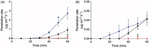
Table 1. Cumulative amount of VX penetrated through human dermatomed skin with or without decontamination.
Figure 2. Agent amount of (A) VX and (B) ethyl methylphosphonic acid (EMPA) on individual tapes. Decontamination with RSDL (green; bottom line), TRSDL (red; bottom line), soapy water (blue; center line) and control without decontamination (black; top line). Values are presented as mean ± SEM (n = 4). *p < 0.05 indicates significantly decreased amounts compared to experiments without decontamination (two-way ANOVA).
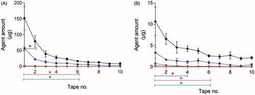
Figure 3. Recovery of skin tissue distribution of VX and ethyl methylphosphonic acid (EMPA) for control experiments without decontamination. The recovery is based on the initial concentration of 95% VX and 5% EMPA exposed on skin. Total amount in 10 tapes (black bar; left), remaining skin (green bar; middle) and penetrated skin (blue bar; right). The VX and EMPA amounts in the remaining skin was calculated by subtraction of the amounts detected in tapes and penetrated skin from the applied amount. Note the two segmented Y-axis. Values are presented as mean ± SEM (n = 4).
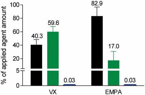
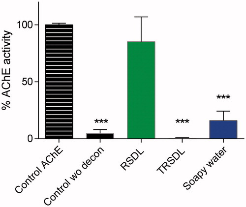
Figure 4. Acetylcholinesterase (AChE) activity measurements by the ChE check mobile for skin surface samples after 10 sets of tape-stripping. Values are presented as mean ± SEM (n = 4). ***p < 0.001 indicates significantly decreased AChE-activity vs the blood control (one-way ANOVA).