Figures & data
Table 1. Demographics table for maternal samples
Figure 1. Manhattan plot of the relationship between maternal and fetal DNA methylation. The x-axis represents the position of each CpG site by chromosome. The y-axis represents the negative log10 of the P-value for the association between maternal and fetal methylation for each CpG site. The red line indicates experiment-wide significance based on a false discovery rate of 5% such that the 5,171 CpG sites above this line are significantly correlated in leukocytes from a mother and her fetus.
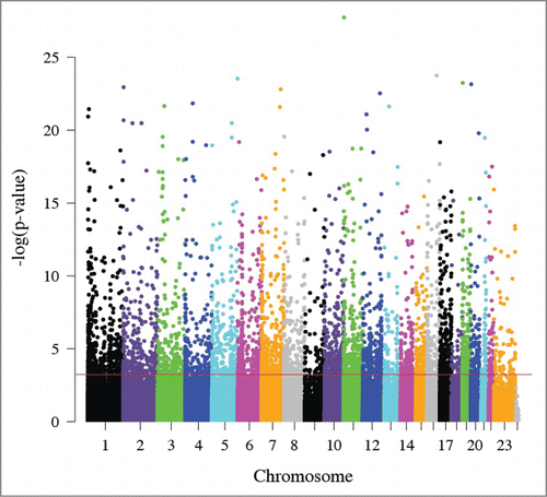
Figure 2. Distribution of maternal DNA methylation for CpG sites. The x-axis represents the standard deviation (SD) of each CpG site's methylation (β) values in maternal leukocytes. The y-axis indicates the proportion of CpG sites in bins determined by SD. Black represents CpG sites that may be attributed to genetic variation while gray represents CpG sites that cannot be attributed to genetic variation. Graph (A) depicts the distribution of correlated CpG sites (n = 5,171). Graph (B) depicts the distribution of all CpG sites (n = 479,808).

Figure 3. Correlated CpG sites and their enrichment in meQTLs. The x-axis represents increasing statistical significance of genotype-methylation associations (cis-meQTLs). The –log10 is used to represent the statistical threshold (α level) used to define meQTLs (i.e., 1 is equivalent to P < 0.1, and 2 is equivalent to P < 0.01, etc.). The y-axis represents the odds ratios comparing the probability that a correlated CpG site associates with a genotype (i.e., is an meQTL) with greater likelihood than an uncorrelated CpG site. The vertical lines represent the confidence interval for each odds ratio.
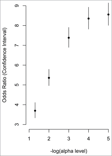
Table 2. Enrichment for correlation analysis of maternal and fetal blood
Table 3. Pathway analysis of CpG sites in genes that correlate in maternal-fetal pairs
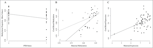
Figure 4. Association of MICB methylation and expression in PTB and TB pairs. (A) Association between cg06284756 in maternal leukocytes and PTB. The x-axis represents PTB status where 0 denotes term birth and 1 denotes PTB. The y-axis represents the methylation levels (β values) for cg06284756. (B) Correlation between maternal methylation (x-axis) and fetal methylation (y-axis) for cg06284756. Open circles represent maternal-fetal pairs that are preterm and closed circles represent pairs that are term. The dashed line represents correlation in PTB pairs, and the solid line represents correlation in term birth (TB) pairs. (C) Correlation between maternal leukocyte MICB expression (x-axis) and fetal leukocyte MICB expression (y-axis).