Figures & data
Figure 1. Experimental design for analyzing the epigenetic effect of high fat diet on male mice sperm cells and on offspring liver gene expression. Sperm cells were prepared from male mice fed a HFD for 10 weeks after weaning. Liver tissues were prepared from males and offspring. Chromatin was prepared and precipitated with antibodies against H3 and H3K4me1, followed by high-throughput sequencing (ChIP-Seq).
Figure 2. Aberrant gene expression in male offspring liver from high fat diet-treated fathers. (A) Real-time PCR analysis in liver from HFD- (red) and LFD-treated (blue) fathers (left panels) and 24-week old male offspring (right panels) for each of the following genes: Acaa1a, Por, Acox1, Fasn, Mt2, and Acaca. For fathers, each bar represents the mean (±SD) of an experiment using 3 animals performed in triplicate. For offspring, each bar represents the mean (±SD) of an experiment using 4 animals performed in triplicate. (B) Real-time PCR analysis in liver from male offspring at 8, 16, and 24 weeks of age from HFD- (red) and LFD-treated (blue) fathers for the genes Mt1, Mt2, Cdkn1a, and Sqle. Each bar represents the result of an experiment performed in triplicate from an individual animal. PCR data were normalized with respect to control Gapdh expression. *P-value < 0.05.
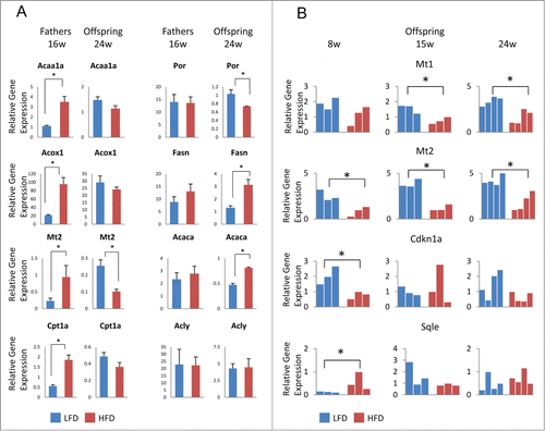
Figure 3. H3 enrichment in HFD- and LFD-treated mice spermatozoa. (A) Distribution of H3 enrichment peaks among gene promoter regions, which include 5 kb upstream and 3 kb downstream the TSS, 5′UTR, 3′UTR, and coding sequence (CDS) in HFD7 vs. LFD8. (B) Heatmap of H3-ChIP signal intensities, normalized for input, around the TSS of protein-coding genes (23,350) in HFD7 and LFD8, at a 5 bp resolution. Profiles show the TSS plus 2 kb of upstream and 2 kb of downstream flanking region. Genes are ranked based on the number of CG sites. (C) Heatmap of H3-enrichment around the TSS of genes encoding microRNAs (1,516) in HFD7 and LFD8 samples. (D) Genome browser view presenting H3-enrichment, normalized to input, in HFD7 and LFD8 samples at genes (black) and CG islands (green bar).
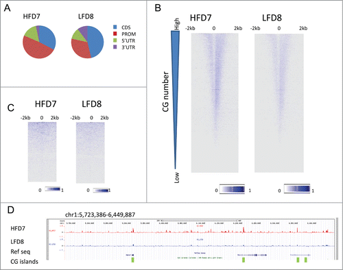
Figure 4. Differential H3 occupancy at regulatory genes in HFD- vs. LFD-treated mice. (A) Heatmap of H3-ChIP signal intensities, normalized to input, around TSS of protein-coding genes (23,350) and 2 kb of flanking sequences for samples HFD7 and LFD8 and their biological replicates, HFD4 and LFD5. Genes are ranked based on H3 occupancy difference at TSS (+500 bp to −500 bp) in HFD7 vs. LFD8, from high- to low-occupancy. (B) GO term analysis of genes highly enriched in H3 (1,074) in HFD samples (HFD7 and HFD4) vs. their control samples (LFD8 and LFD5). (C) Genome browser view presenting H3-enrichment, normalized to input, for sample sets HFD7 (red) and LFD8 (blue) and biological replicates HFD4 (red) and LFD5 (blue) at selected genes involved in transcription regulation.
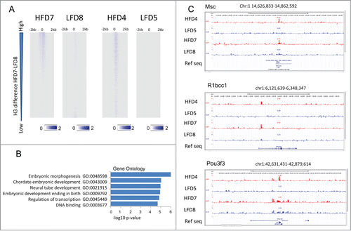
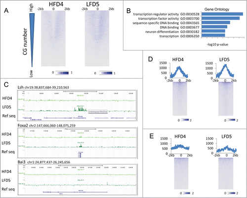
Figure 5. H3K4me1 enrichment at TSS and enhancer. (A) Heatmap of H3K4me1-ChIP signal intensities, normalized to input, around TSS of protein-coding genes (23,350) at a 5-bp resolution in HFD4 vs. LFD5 spermatozoa samples. Profiles show TSS and 2 kb of upstream and 2 kb of downstream flanking region. Genes were ranked based on the number of CG sites. (B) GO term analysis of the top 5% of genes highly enriched in H3K4me1 (1,169) in LFD5 vs. HFD4 samples. (C) Genome browser view representing H3K4me1 enrichment, normalized to input, for HFD4 (dark green) and LFD5 (light green) at selected genes encoding transcriptional regulators. (D) Heatmap of H3K4me1-ChIP signal intensities at enhancers (9,452) plus 2 kb of upstream and downstream flanking sequences, normalized to input, at a 25-bp resolution in HFD4 vs. LFD5 spermatozoa samples. Sequences were sorted based on highest enrichment in HFD4. Line graphs on top of heatmaps represents the sum of H3K4me1 enrichment profiles at each position. (E) Heatmap of H3K4me1-ChIP signal intensities at enhancers (8,701) in liver samples.