Figures & data
Table 1. Characteristics of study participants.
Figure 1. Differentially methylated CpGs between HBV-HCC vs. HCV-HCC. (A) Principal component analysis of the methylation data was plotted using the first 2 principal components (PC1 = 67.2% and PC2 = 9.04%). Each dot represents a sample (red for HBV-HCC and blue for HCV-HCC). (B) Unsupervised hierarchical clustering of β-values for differentially methylated loci. Red and blue blocks on the top of the maps represent HBV-HCC (n = 10) and HCV-HCC (n = 13), respectively. (C) Mean methylation level for all 7 differentially-methylated loci among HBV-HCC (left) and HCV-HCC (right). (D) The top IPA network involving differentially methylated genes. Red and blue genes indicate differentially methylated genes and the connected cancer related genes, respectively. (E) The top associated cellular functions with numbers of differentially-methylated genes.
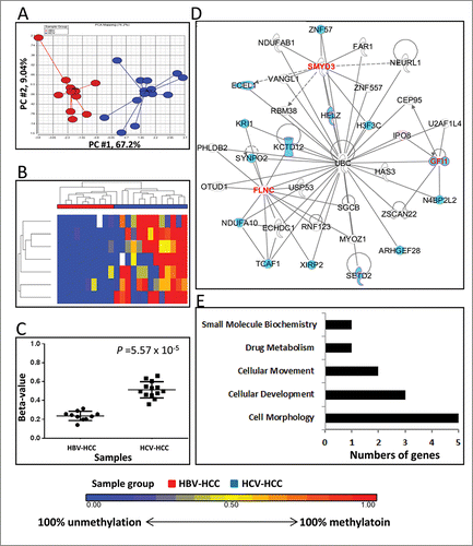
Figure 2. Differentially methylated CpGs between HBV-HCC vs. NIV-HCC. (A) Principal component analysis of the methylation data was plotted using the first 2 principal components (PC1 = 68.4% and PC2 = 6.55%). Each dot represents a sample (red for HBV-HCC and green for NIV-HCC). (B) Unsupervised hierarchical clustering of β-values for differentially methylated loci. Red and green blocks on the top of the maps represent HBV-HCC (n = 10) and NIV-HCC (n = 10), respectively. (C) Mean methylation level for all 7 differentially-methylated loci among HBV-HCC (left) and NIV-HCC (right). (D) The top IPA network involving differentially methylated genes. Red and blue genes indicate differentially methylated genes and the connected cancer related genes, respectively. (E) The top associated cellular functions with numbers of differentially-methylated genes.
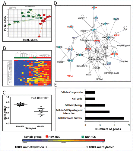
Figure 3. Differentially methylated CpGs between HCV-HCC vs. NIV-HCC. (A) Principal component analysis of the methylation data was plotted using the first 2 principal components (PC1 = 64% and PC2 = 6.65%). Each dot represents a sample (blue for HCV-HCC and green for NIV-HCC). (B) Unsupervised hierarchical clustering of β-values for differentially methylated loci. Blue and green blocks on the top of the maps represent HCV-HCC (n = 13) and NIV-HCC (n = 10), respectively. (C) Mean methylation level for all 7 differentially-methylated loci among HCV-HCC (left) and NIV-HCC (right). (D) The top IPA network involving differentially methylated genes. Red and blue genes indicate differentially methylated genes and connected cancer related genes, respectively. (E) The top associated cellular functions with numbers of differentially-methylated genes.
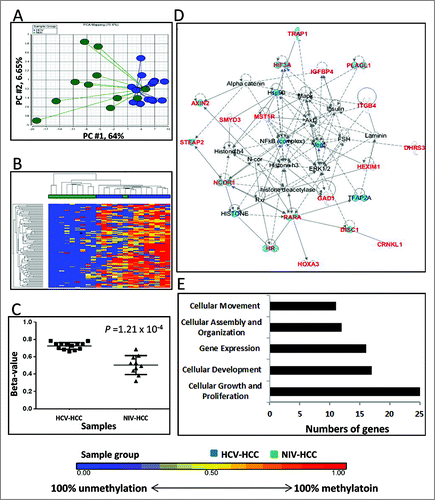
Table 2. Correlations of DM loci and gene expression.
Figure 4. Association between differentially methylated loci by virus infection and other HCC risk factors. Mann-Whitney rank test was used for alcohol consumption (ever vs. never), cirrhosis (yes vs. no), diabetes (yes vs. no), and smoking (yes vs. no) and Spearman correlation was used for BMI (continuous variable).
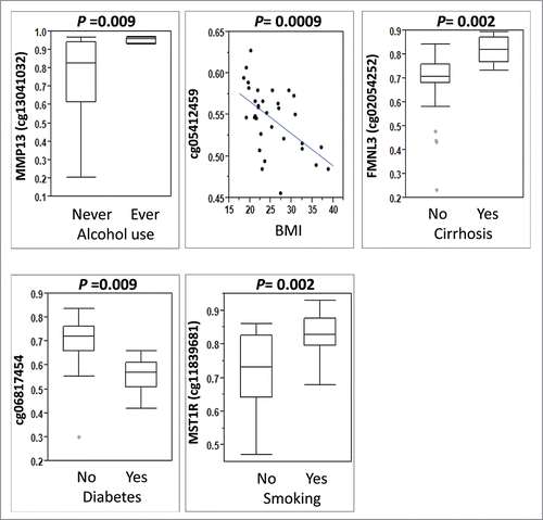
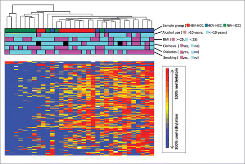
Figure 5. Unsupervised hierarchical clustering of β-values for all differentially methylated loci identified by different viral infection. Unsupervised hierarchical clustering of β-values for DM loci (rows) in 33 HCC samples (columns). Red, blue, green blocks on the top of the maps represent HBV-HCC (n = 10), HCV-HCC (n = 13) and NIV-HCC (n = 10), respectively. The clinical characteristics of the samples including alcohol use, BMI, cirrhosis, diabetes, and smoking were represented as other blocks below sample group.