Figures & data
Table 1. Summary of study participants' characteristics.
Figure 1. Odds ratios for having an SGA or LGA vs. AGA infant in association with levels of extracellular microRNAs (exmiRNAs) in maternal serum. Odds ratios are adjusted for gestational age and infant's sex; -log10 (P Value) indicates transformed P values of the association between exmiRNAs and SGA or LGA. Odds ratios between SGA and AGA are shown in white and between LGA and AGA in gray.
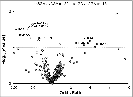
Figure 2. Associations between extracellular microRNAs (exmiRNAs) in maternal serum and birth weight-for-gestational age z-score (n = 100). Volcano plot showing the association between serum exmiRNAs at second trimester and birth weight-for-gestational age z-score; estimates are adjusted for maternal age, gestational age, body mass index, parity, and infant's sex; -log10 (P value) indicates transformed P values of the association between exmiRNAs and birth weight-for-gestational age z-score.
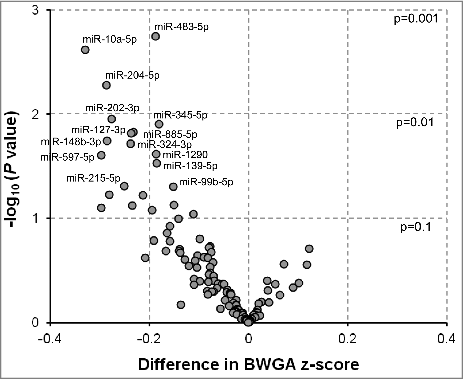
Figure 3. Pathway analysis illustrating experimentally validated mRNA targets of the most associated exmiRNAs with fetal growth. Biological relevance with functions and diseases related to fetal growth is also identified by the Ingenuity Pathway Analysis®. Direct relationships are represented with solid lines and indirect relationships with dotted lines.
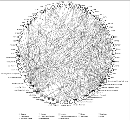
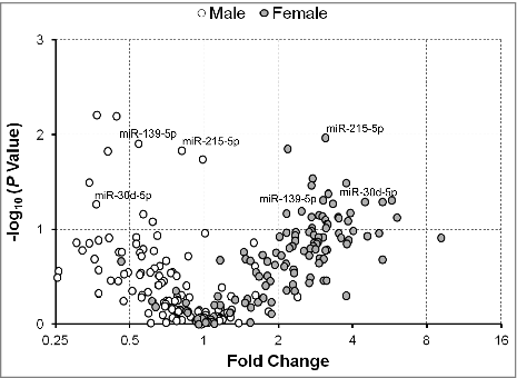
Figure 4. Fold change of serum extracellular microRNAs (exmiRNAs) between mothers of small- and appropriate-for-gestational age infants by gender. Volcano plot showing the fold change of all tested serum exmiRNAs at second trimester from mothers of male (white) and female (gray) SGA vs. AGA infants. *Denotes exmiRNAs in female mother-infant pairs.
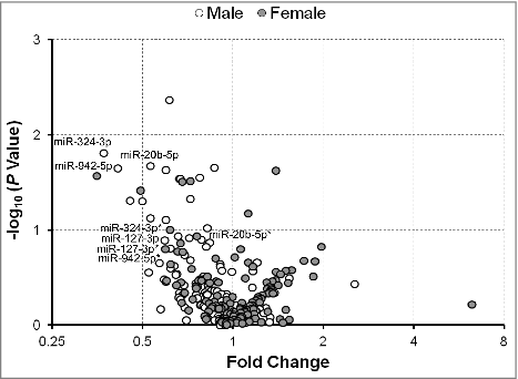
Figure 5. Fold change of serum extracellular microRNAs (exmiRNAs) between mothers of large- and appropriate-for-gestational age infants by gender. Volcano plot showing the fold change of all tested serum exmiRNAs at second trimester from mothers of male (white) and female (gray) LGA vs. AGA infants.