Figures & data
Figure 1. Tet enzyme gene expression is highest at birth. Expression of Tet1, Tet2 and Tet3 mRNA at postnatal days 1, 7, 25, and 60 in a) the preoptic area of the hypothalamus (POA); b) the ventromedial nucleus of the hypothalamus (VMH); and c) the hippocampus. The expression of all three Tet enzymes was highest in newborns, and abruptly decreased by day 7. Different lower-case letters indicate statistically significant differences (P < 0.05). Data are mean ± SEM. N = 19–25 mice per age.
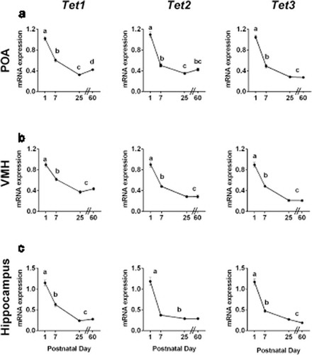
Figure 2. Canonical Dnmt gene expression is highest during neonatal life, whereas expression of the co-activator Dnmt3l increases during postnatal life. Dnmt1, Dnmt3a, Dnmt3b and Dnmt3l mRNA expression at postnatal days 1, 7, 25 and 60 in a) the preoptic area of the hypothalamus (POA); b) the ventromedial nucleus of the hypothalamus (VMH); and c) the hippocampus. The expression of Dnmt1 and Dnmt3b was highest on P1, then dropped to lower levels by P7, whereas expression of Dnmt3a remained elevated throughout the first postnatal week in all brain regions examined. Expression of Dnmt3l increased 6- to 10-fold between postnatal days 1 and 60 in all brain regions. Different letters indicate significant differences. Data are mean ± SEM. N = 19–20 mice per age.
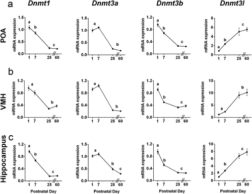
Figure 3. Sex differences in Tet gene expression are observed during neonatal life. In the preoptic area of the hypothalamus (POA), Tet2 and Tet3 expression are higher in males than in females on postnatal day (P) 7. Neonatal testosterone treatment partially masculinized Tet3 expression in females, but did not affect the expression of Tet2. N = 5–7 per group. In the ventromedial nucleus of the hypothalamus (VMH), expression of Tet1 and Tet2 is higher in males on P1, and neonatal testosterone treatment of females did not eliminate the sex difference. N = 6 in each female group, N = 13 males. In the hippocampus, females have marginally higher expression of each Tet enzyme on P1; across all three Tet enzymes, expression was significantly higher in females than in males (P < 0.01). Expression in females treated neonatally with testosterone was intermediate, and not significantly different from that of control males or females. N = 6 in each female group, N = 13 in males. #P < 0.1, *P < 0.05; **P < 0.01; ****P < 0.0001. Data are mean ± SEM.
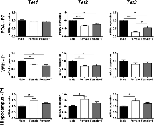
Figure 4. Females have higher expression of Dnmt1 and Dnmt3l in the preoptic area of the hypothalamus (POA) at postnatal day (P) 7. Dnmt1 expression in females treated with testosterone was intermediate and not significantly different from that of control males or females. *P < 0.05. Data are mean ± SEM. N = 5 vehicle females, N = 7 males and females + testosterone.
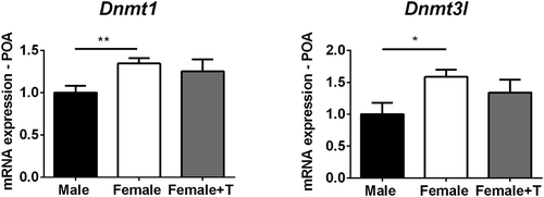
Figure 5. Tet enzyme activity is elevated at birth. a) The activity of demethylating (Tet) enzymes was two-to three-fold higher on postnatal day (P) 1 than on P25 in both the hypothalamus and hippocampus. b) There was a trend for higher methylating (Dnmt) activity in the hypothalamus on P1 compared to P25. In the hippocampus, elevated Dnmt activity on postnatal day 1 was seen in females only. Different letters indicate significant differences. **P < 0.01; ***P < 0.001. Data are mean ± SEM. N = 3 for P1 male and female groups, N = 6 for P25 male and female groups.
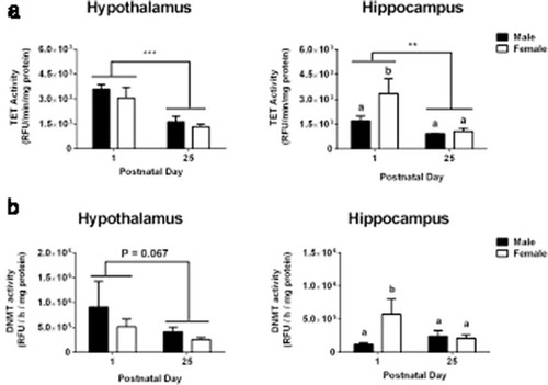
Figure 6. Global levels of 5-methylcytosine (5mC) in the preoptic area of the hypothalamus (POA) increase across development. Global 5mC significantly increased between postnatal day 7 and 25 (P < 0.001). A sex difference was found at postnatal day 7, with two-fold higher global methylation in females than in males. Neonatal testosterone treatment of females did not masculinize global methylation. Different letters indicate significant differences. *P < 0.05 and ***P < 0.001, compared to males on postnatal day 7. Data are mean ± SEM. N = 6 in all groups.
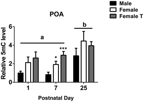
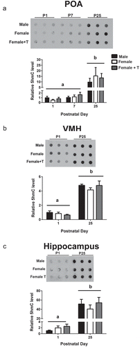
Figure 7. Global 5-hydroxymethylcytosine (5hmC) accumulates across development. Representative dot blots and quantification illustrate that hydroxymethylation significantly increases between postnatal days 1 and 25 in a) the preoptic area of the hypothalamus (POA); b) the ventromedial nucleus of the hypothalamus (VMH); and c) the hippocampus (P < 0.001 in all cases). Different letters indicate significant differences. Data are mean ± SEM. N = 6 in all groups.