Figures & data
Table 1. Clinical and biochemical characteristics of control and PCOS pregnant women with (PCOS+M) and without metformin (PCOS)
Table 2. Clinical characteristics of control and PCOS infants (daughters and sons) born to control and PCOS mothers treated (PCOS+M) and non-treated (PCOS) with metformin during pregnancy
Figure 1. Methylation levels (β-value) in the promoter regions of the leptin receptor (LEPR) (a), leptin (LEP) (b), adiponectin receptor 2 (ADIPOR2) (c) and androgen receptor (AR) genes (d) in daughters of control (control, n = 12), PCOS women (PCOS, n = 6) and PCOS women treated with metformin during pregnancy (PCOS+M, n = 6). Data are shown as median ± SEM. Dots indicate the cases in each group. Differences were calculated by one-way ANOVA followed by Bonferroni test or Kruskal-Wallis test followed by Dunn test. aP < 0.05 between control and PCOS; bP < 0.05 between control and PCOS+M; cP < 0.05 between PCOS and PCOS+M
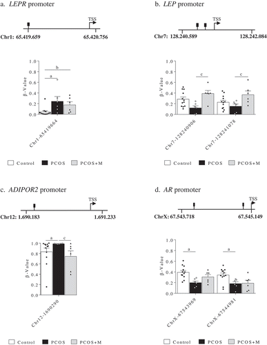
Figure 2. Methylation levels (β-value) in the promoter regions of the leptin (LEP) (a), antimüllerian hormone (AMH) (b) and androgen receptor (AR) genes (c) in sons of control (control, n = 12), PCOS women (PCOS, n = 6) and PCOS women treated with metformin during pregnancy (PCOS+M, n = 6). Data are shown as median ± SEM. Dots indicate the cases in each group. Differences were calculated by one-way ANOVA followed by Bonferroni test or Kruskal-Wallis test followed by Dunn test. aP < 0.05 between control and PCOS; bP < 0.05 between control and PCOS+M; cP < 0.05 between PCOS and PCOS+M
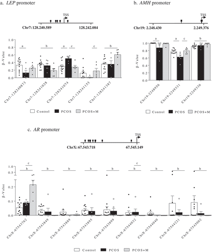
Figure 3. Z-Score of the promoter regions of LEP, LEPR, ADIPOQ, ADIPOR1, ADIPOR2, AMH and AR in daughters and sons of control (control, n = 12/12), PCOS women (PCOS, n = 6/6) and PCOS women treated with metformin during pregnancy (PCOS+M, n = 6/6). Data are shown as median ± SEM. Dots indicate the cases in each group. Differences were calculated by Kruskal-Wallis test followed by Dunn test. aP < 0.05 between control and PCOS; bP < 0.05 between control and PCOS+M
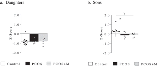
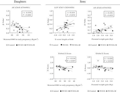
Figure 4. Correlation analysis between the methylation levels (β-value) of the CpG sites of the promoter regions of the AR (chromosome X, ChrX), and LEP (chromosome 7, Chr7) genes (and Z-Score of the analysed promoter regions), and the maternal characteristics and anthropometric parameters at birth and postnatal age in daughters and sons. Dots indicate the cases in each group. The associations between variables were calculated by Spearman’s rank correlation analysis. P < 0.05 was considered as significant level