Figures & data
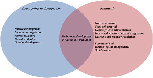
Figure 1. TET-driven demethylation on different substrates (RNA/DNA) in mammals and D. melanogaster. (a) TET in mammals and D. melanogaster demethylates the DNA on cytosines and adenosines, respectively. RNA demethylation is also carried out by TET in D. melanogaster; however, it has only been investigated in vitro in mammals. (b) A schematic representation of active DNA demethylation by TET, TDG (thymidine-DNA-glycosylase) and BER (base excision repair)-mediated pathways
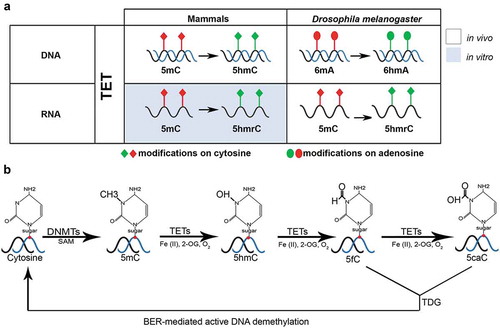
Table 1. The function of 6mA and 5hmrC modification in different multicellular organisms
Figure 2. Comparison of human and D. melanogaster TET protein structure and expression. (a). Three TET proteins are found in humans in comparison to one TET (dTet) homologue in D. melanogaster. All share a conserved cysteine-rich domain (dark blue colour) and a DSBH (Double Stranded Beta-Helix) domain (dark grey colour). TET2 lacks the CXXC DNA binding domain (light blue colour). The Fe(II)-binding site (red colour) and 2-oxoglutarate binding site (yellow colour) are both located within the catalytic domain. (b). An overview of TET expressing tissues in humans and D. melanogaster.
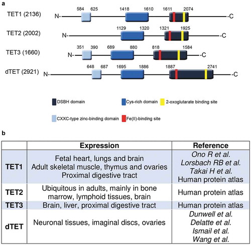
Figure 3. Comparison between the function of TET proteins in D. melanogaster and mammals