Figures & data
Figure 1. Potential redox of glutathione and vitamin C concentrations depending of nutritive modalities
ON: animals fed regular food; PN-IL-LE: animals fed exclusively with total parenteral nutrition (PN) compounded with Intralipid without photoprotection; PN-IL-LP: photoprotected PN compounded with Intralipid; PN-SF-LE: PN compounded with SMOFLipid without photoprotection; PN-SF-LP: photoprotected PN compounded with SMOFLipid. Panel A: The redox potential was more oxidized in the PN groups than in the ON group and more in SF groups. Panel B: Levels of vitamin C (ascorbate + dehydroascorbate) were higher in the photoprotected PN groups. Data are expressed as mean ± s.e.m., n = 6–8 per group. The bars show the statistical comparisons. The absence of a symbol on the bar means that p > 0.05. *: p < 0.05; ***: p < 0.001.
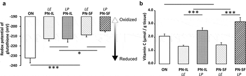
Table 1. Hepatic GSH and GSSG levels depending on nutritive modalities
Table 2. Characteristics of lipid emulsions
Figure 2. DNA methylation depending of nutritive modalities
ON: animals fed regular food; PN-IL-LE: animals fed exclusively with total parenteral nutrition (PN) compounded with Intralipid without photoprotection; PN-IL-LP: photoprotected PN compounded with Intralipid; PN-SF-LE: PN compounded with SMOFLipid without photoprotection; PN-SF-LP: photoprotected PN compounded with SMOFLipid. Panel A: The hepatic level of 5-MedCyd (5ʹ-methyl-2ʹ-deoxycytidine) increased in the PN groups. The SF effect did not reach the significance level of p = 0.05. Panel B: the DNA methylation values were significantly associated with the redox value of glutathione (y = 0.16 pmol •μg DNA−1•mV−1 •x + 41 pmol •μg DNA−1; r2 = 0.51, p < 0.0001). Open triangle: ON; open circle: PN-IL-LE; open square: PN-SF-LE; black circle: PN-IL-LP; black square: PN-SF-LP. Data are expressed as mean ± s.e.m., n = 6 per group. The bars show the statistical comparisons. The absence of a symbol on the bar means that p > 0.05. **: p < 0.01.
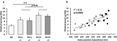
Figure 3. DNA methylation activity, levels of SAM and SAH depending of nutritive modalities
ON: animals fed regular food; PN-IL-LE: animals fed exclusively with total parenteral nutrition (PN) compounded with Intralipid without photoprotection; PN-IL-LP: photoprotected PN compounded with Intralipid; PN-SF-LE: PN compounded with SMOFLipid without photoprotection; PN-SF-LP: photoprotected PN compounded with SMOFLipid. Panel A: The activity of DNMTs was higher in PN groups than in ON group. Panel B: The hepatic concentration of SAM was higher in the PN groups and more in the IL groups. Panel C: The concentration of SAH was lower in SF groups. Panel D: Relationship between the ratio SAM on SAH and the redox potential of glutathione (y = 0.045 mV−1 • x – 11.84; r2 = 0.36, p < 0.001). Open triangle: ON; open circle: PN-IL-LE; open square: PN-SF-LE; black circle: PN-IL-LP; black square: PN-SF-LP. Data are expressed as mean ± s.e.m., n = 4–6 per group. The bars show the statistical comparisons. The absence of a symbol on the bar means that p > 0.05. **: p < 0.01.
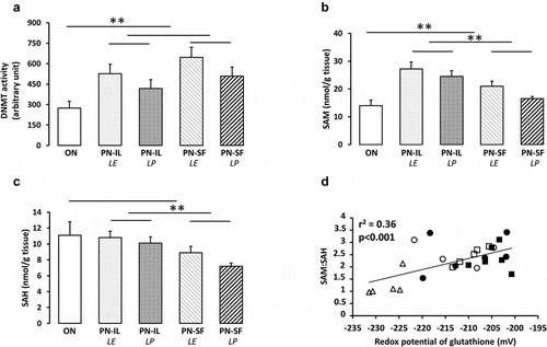
Figure 4. DNMT1 and DNMT3b protein levels depending of nutritive modalities
ON: animals fed regular food; PN-IL-LE: animals fed exclusively with total parenteral nutrition (PN) compounded with Intralipid without photoprotection; PN-IL-LP: photoprotected PN compounded with Intralipid; PN-SF-LE: PN compounded with SMOFLipid without photoprotection; PN-SF-LP: photoprotected PN compounded with SMOFLipid. Panel A and B: DNMT1 protein levels were lower in PN groups compared to ON groups. Exposure to light was significant only in the PN-IL groups. Panel C and D: Western blot of DNMT3b protein levels showed no statistical difference between groups. Data are expressed as mean ± s.e.m., n = 7–8 per group. The bars show the statistical comparisons. The absence of a symbol on the bar means that p > 0.05. *: p < 0.05; **: p < 0.01.
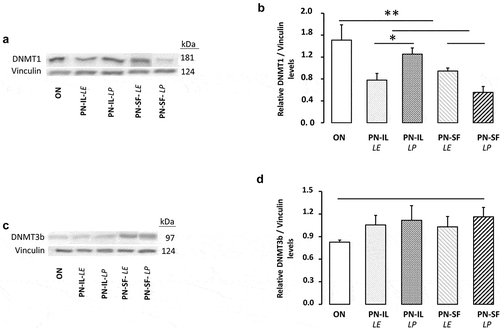
Figure 5. DNMT3a protein levels depending of nutritive modalities
ON: animals fed regular food; PN-IL-LE: animals fed exclusively with total parenteral nutrition (PN) compounded with Intralipid without photoprotection; PN-IL-LP: photoprotected PN compounded with Intralipid; PN-SF-LE: PN compounded with SMOFLipid without photoprotection; PN-SF-LP: photoprotected PN compounded with SMOFLipid. Panel A: Western blot of DNMT3a isoforms. Panel B and C: DNMT3a1 and DNMT3a2 protein levels were higher in PN groups compared to ON groups. PN-SF resulted in significant increased in DNMT3a1 protein expression compared to PN-IL and higher in the photoprotected PN groups. Data are expressed as mean ± s.e.m., n = 6 per group. The bars show the statistical comparisons. The absence of a symbol on the bar means that p > 0.05. *: p < 0.05; **: p < 0.01; ***: p < 0.001.
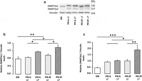
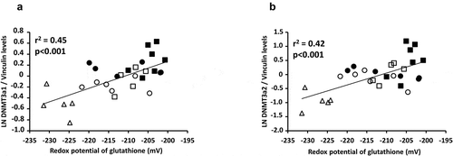
Figure 6. Relationships between DNMT3a and redox potential of glutathione
Panel A: The relationship between the natural logarithm (LN) value of DNMT3a1 and redox potential was significant (y = 0.025 pixel mV−1 x + 5.19 pixel; r2 = 0.45; p < 0.001). Panel B: the relationship between the natural logarithm (LN) value of DNMT3a2 and the redox potential was significant (y = 0.043 pixel mV−1 + 8.98 pixel; r2 = 0.42; p < 0.001). Open triangle: ON; open circle: PN-IL-LE; open square: PN-SF-LE; black circle: PN-IL-LP; black square: PN-SF-LP.