
Figures & data
Figure 1. Cyp2a5 expression in liver of PSE and control in E17.5, D3 mice and adult offspring
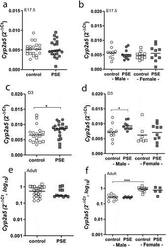
Figure 2. Sex-dependent Cyp2a5 promoter methylation across three time points (E17.5, D3 mice and adult offspring). DNA was isolated from whole liver of foetuses (E17.5, a), neonatal offspring (D3, b) and adult offspring (c) in PSE (closed symbols) and control groups (open symbols). DNA was subjected to bisulphite sequencing-based methylation analysis of the Cyp2a5 promoter region and the percentage of DNA methylation was assessed. Data of the 6 targeted CpG-sites are presented per sex and exposure as individual values with median as a horizontal line. CpG-site annotations are relative to the ATG start codon. If not stated otherwise, the comparison of shown groups was not significant. *p ≤ 0.05, ** p ≤ 0.01, ***p ≤ 0.001 (Mann-Whitney U-test). Circle (○) symbol(s) = male, square (□) symbol(s) = female
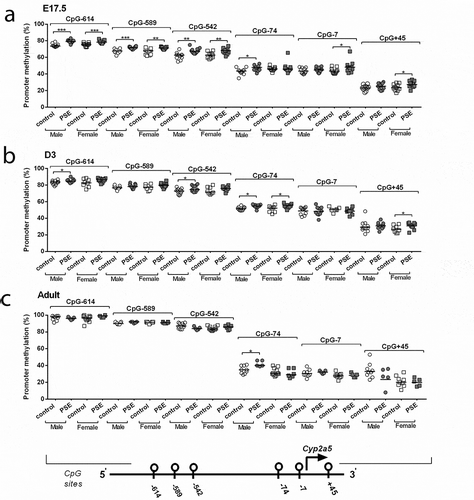
Table 1. Sequences of primers used in bisulphite-based methylation analysis
Table 2. Correlations between Cyp2a5 mRNA expression and promoter methylation in foetal (E17.5), neonatal (D3) and adult offspring liver
Figure 3. Time points comparisons of sex-dependent Cyp2a5 promoter methylation status in PSE and control groups. The ANOVA test was used to do the comparison analysis among the foetal stage (E17.5), neonatal period (D3 means three days after birth) and adulthood (D140 means 140 days later after birth). P-values<0.0001 were detected by ANOVA test in all of CpG sites over three time points. Mann–Whitney U-test was used to test the comparisons between two time points in control (open symbols) and PSE (closed symbols) groups for 6 promoter methylation CpG sites. Data are represented as mean± SEM; CpG-site annotations are relative to the ATG start codon. *p ≤ 0.05, *** p ≤ 0.001 (Mann-Whitney U-test). Circle (○) symbol(s) = male, square (□) symbol(s) = female
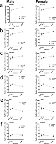
Figure 4. Optimization and the cotinine formation levels of in vitro nicotine metabolism in E17.5, D3 mice and adult offspring. Cotinine formation of nicotine metabolism of foetuses (E17.5), three-day-old offspring (D3) and adult offspring in PSE and control groups. The nicotine metabolism was measured by HPLC-L as described in the material and methods section. The assay was optimized with respect to incubation time (a) and cytosolic protein concentration (b). Arrows at the X-axis indicate conditions used for actual experiments. C presents the HPLC chromatogram separation of a blank sample containing the standards nicotine and cotinine and the cotinine was produced in liver microsomes of PSE male neonatal offspring. D presents the cotinine analysis by LCMS. Cotinine, corresponding with m/z 177 was detected both in standard cotinine and in the microsome sample. The levels of cotinine formation were measured in foetal (e), three-day-old offspring (f) and adult offspring (g) of both groups. Circle (○) symbol(s) = male, square (□) symbol(s) = female. Data are shown as individual values. If not stated otherwise, the difference between groups was not significant. *p ≤ 0.05, ** p ≤ 0.01 (Mann-Whitney U-test). Open symbol(s) = control group, closed symbol(s) = PSE
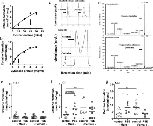
Table 3. Correlations between cotinine levels, Cyp2a5 mRNA expression and promoter methylation in foetal (E17.5), neonatal (D3) and adult offspring liver