Figures & data
Table 1. Characteristics of the selected mother-newborn pairs, categorized by exposure group
Figure 1. Mean mitochondrial DNA methylation levels as determined by bisulphite pyrosequencing (n = 20/group). The horizontal line is equivalent to the median. The lower and upper hinges represent the first and third quartiles. The upper whisker extends from the hinge to the largest value no further than 1.5 * IQR (= interquartile range) from the hinge, whereas the lower whisker extends from the hinge to the smallest value no further than 1.5 * IQR of the hinge. Data outside this range is represented as dots
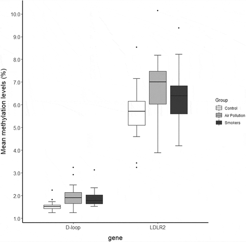
Table 2. Differences in mtDNA methylation levels between exposure groups as determined by linear mixed models
Table 3. Association between placental mtDNA methylation levels and newborn birth weight
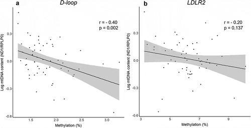
Figure 2. Correlation between mitochondrial DNA content and absolute mitochondrial DNA methylation levels in placenta tissue for the D-loop (a) and LDLR2 (b) regions