Figures & data
Table 1. The primer sequence used in this study.
Figure 1. TET1 expression was downregulated in the cervical cancer tissues. (a) The expression of TET1 in the cervical cancer tissues with different degrees of differentiation; (b) The TET1 expression in cervical cancer and normal cervical tissues; (c) TCGA data indicating that TET1 was downregulated in cervical cancer.
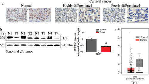
Figure 2. TET1 knockdown promoted the migration and invasion of cervical cancer cells.
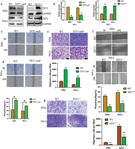
Figure 3. TET1 reduces the proliferation, clonal formation, and tumorigenesis of cervical cancer cells.
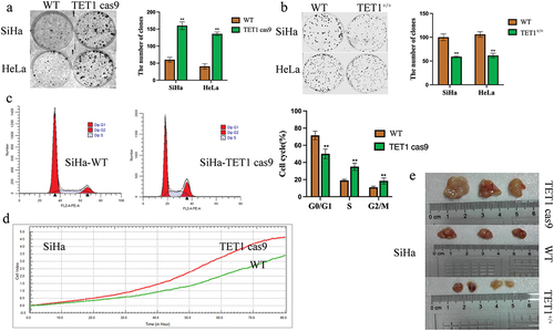
Figure 4. Effect of TET1 on epithelial-mesenchymal transformation (EMT) of cervical cancer SiHa cells.
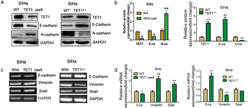
Figure 5. Effect of TET1 on the biological functions of cervical cancer cells.
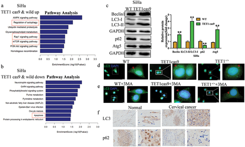
Figure 6. Effects of autophagy on TET1-mediated biological changes in cervical cancer cells.
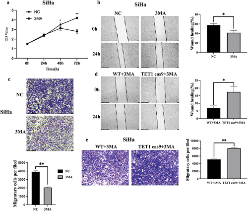
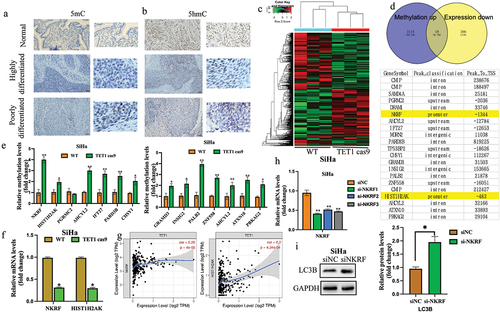
Figure 7. Effect of TET1 on the gene methylation level and mRNA expression.