Figures & data
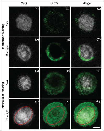
Figure 1. Subcellular localization of Atcry2 in insect cells by immunofluorescence staining and confocal microscopy.Sf21 cells stably expressing Atcry2 were fixed with paraformaldehyde, permeabilized with Triton X100 (A) to (F) or not (G) to (L), incubated with a rabbit polyclonal anti-Atcry2 antibody and an Alexa 488-conjugated anti-rabbit secondary antibody, DNA were stained with Dapi. Cells were observed with an inverted Leica TCS SP5 microscope. Images (A) to (F) show projection of optical sections, scale bar 10 μm. Images (G) to (L) show single confocal z-section that cross the nucleus, scale bar 10 μm.

Figure 2. Production and subcellular localization of ROS by Sf21 CRY2 exposed to blue light.Living Sf21 stably expressing CRY2 were exposed to dark or blue light, treated with DCFH-DA [5-(and-6)-chloromethyl-2′,7′-dichlorofluorecein diacetate] and viewed by an inverted Leica TCS SP5 microscope. Images show single confocal z section that cross the nucleus. Intense ROS staining can be seen inside the nucleus (N) whose membrane can be clearly observed by differential interference contrast (D. I. C.). Scale bars 10 μm. Methods for AtCry2 expression, immunohistochemical staining, and fluorescence staining for ROS are described in our original paper.Citation3 Polyclonal antibody used for cry2 detection has been raised to the C-terminal domain as used previously. Citation6
![Figure 2. Production and subcellular localization of ROS by Sf21 CRY2 exposed to blue light.Living Sf21 stably expressing CRY2 were exposed to dark or blue light, treated with DCFH-DA [5-(and-6)-chloromethyl-2′,7′-dichlorofluorecein diacetate] and viewed by an inverted Leica TCS SP5 microscope. Images show single confocal z section that cross the nucleus. Intense ROS staining can be seen inside the nucleus (N) whose membrane can be clearly observed by differential interference contrast (D. I. C.). Scale bars 10 μm. Methods for AtCry2 expression, immunohistochemical staining, and fluorescence staining for ROS are described in our original paper.Citation3 Polyclonal antibody used for cry2 detection has been raised to the C-terminal domain as used previously. Citation6](/cms/asset/ceeaa652-8245-4361-8663-43a939a40d5f/kpsb_a_1042647_f0002_oc.gif)
