1,239
Views
11
CrossRef citations to date
0
Altmetric
Short Communication

Daurichromenic acid and grifolic acid: Phytotoxic meroterpenoids that induce cell death in cell culture of their producer Rhododendron dauricum

ORCID Icon, &
Article: e1422463 | Received 10 Nov 2017, Accepted 21 Dec 2017, Published online: 16 Jan 2018

Figures & data

Figure 1. Biosynthesis of daurichromenic acid (DCA). DCA is synthesized by DCA synthase via stereoselective oxidocyclization of the farnesyl moiety of grifolic acid. Two electrons from the substrate are abstracted by enzyme-bound flavin adenine dinucleotide (FAD), and then transferred to molecular oxygen to release H2O2 as the by-product, as described previously.Citation4

Figure 1. Biosynthesis of daurichromenic acid (DCA). DCA is synthesized by DCA synthase via stereoselective oxidocyclization of the farnesyl moiety of grifolic acid. Two electrons from the substrate are abstracted by enzyme-bound flavin adenine dinucleotide (FAD), and then transferred to molecular oxygen to release H2O2 as the by-product, as described previously.Citation4

Figure 2. Phytotoxic effects of daurichromenic acid (DCA) and grifolic acid on R. dauricum cells. (A) Trypan blue staining to evaluate cell viability. Seven-day-old cell cultures were incubated with 100 µM of each indicated compound for 24 h, then stained with trypan blue. Control cells were treated with the solvent dimethyl sulfoxide (DMSO) only. White Bar, 100 µm. Inserted images are the fluorescent detection of nuclei after staining with 4′,6-diamidino-2-phenylindole (DAPI). Yellow bar, 10 µm. (B) Agarose gel electrophoresis of genomic DNA from R. dauricum cells treated as in A. DNA was separated on a 2% agarose gel and stained with ethidium bromide. Lane M, marker DNA with the indicated sizes; lane 1, control cells; lane 2, DCA-treated cells; lane 3, grifolic acid-treated cells; lane 4, orsellinic acid-treated cells; and lane 5, camptothecin-treated cells.
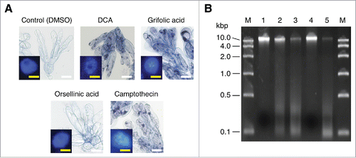
Figure 2. Phytotoxic effects of daurichromenic acid (DCA) and grifolic acid on R. dauricum cells. (A) Trypan blue staining to evaluate cell viability. Seven-day-old cell cultures were incubated with 100 µM of each indicated compound for 24 h, then stained with trypan blue. Control cells were treated with the solvent dimethyl sulfoxide (DMSO) only. White Bar, 100 µm. Inserted images are the fluorescent detection of nuclei after staining with 4′,6-diamidino-2-phenylindole (DAPI). Yellow bar, 10 µm. (B) Agarose gel electrophoresis of genomic DNA from R. dauricum cells treated as in A. DNA was separated on a 2% agarose gel and stained with ethidium bromide. Lane M, marker DNA with the indicated sizes; lane 1, control cells; lane 2, DCA-treated cells; lane 3, grifolic acid-treated cells; lane 4, orsellinic acid-treated cells; and lane 5, camptothecin-treated cells.
Supplemental material

Reprints and Corporate Permissions

Please note: Selecting permissions does not provide access to the full text of the article, please see our help page How do I view content?

To request a reprint or corporate permissions for this article, please click on the relevant link below:

Academic Permissions

Please note: Selecting permissions does not provide access to the full text of the article, please see our help page How do I view content?

Obtain permissions instantly via Rightslink by clicking on the button below:

If you are unable to obtain permissions via Rightslink, please complete and submit this Permissions form. For more information, please visit our Permissions help page.