Figures & data
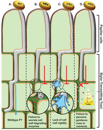
Figure 1. Potential models for AtOFT1 behavior during pollen tube penetration.
Illustration of proposed mechanisms for the oft1 mutant pollen tube penetration defect. A, Col-0 (WT) PT rapidly traversing the transmitting tract towards the ovary, while oft1 mutant PTs (B-D) exhibit slower penetration rates through these tissues, which most often results in arrested growth. B, The oft1 PT may fail to secrete or activate female tissue cell wall degrading enzymes, thus making passage through these tissues arduous. C, oft1 PT potentially lacks a cell wall rigidifying component that aids in penetrating though the female tissue layers during fertilization. D, oft1 PT potentially lacks a functional receptor that is necessary for the PT to respond to positional guidance cues within the stylar transmitting tract (TT) or to metabolize the nutrients supplied by the TT ECM. Borders around cells are cuticle (purple), cell wall (green), and plasma membrane (orange). The location of the papillar cells and the stylar transmitting tract are indicated.