Figures & data
Table 1. Analysis of embryo sac and early seed development in cleared ovaries of apomictic Hieracium praealtum (4x-1 = 35). Total ovules scored represent the number of ovules in five main categories, excluding subcategories.
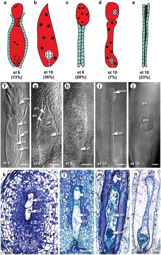
Figure 1. Representative examples of embryo sac and embryo formation during aposporous development in H. praealtum.
(A) to (E) Cartoons depicting frequent types of embryo sacs observed in ovules of H. praealtum. (A) Embryo sac with enlarged AI cell at the top. (B) Single embryo sac. (C) Collapsed endothelium with embryo sac at the chalazal end. (D) Twin embryo sacs with developing embryos. (E) Aborted embryo sac resulting in collapsed endothelium. (F) to (J) Differential interference contrast images of off-type embryo sacs. (F) Two embryo sacs with embryo developing in the sac at the chalazal end, an AI is present within the top of the micropylar embryo sac. (G) An embryo sac developing endosperm but no embryo, in the top of this embryo sac is another. Bars = 50 µm. (H) Endosperm development without embryo in a chalazal embryo sac. (I) Collapsed endothelium with developing embryo at the chalazal end. (J) Lateral embryo development mid embryo sac. Arrowheads point toward structures within embryo sacs. Bars = 100 µm. (K) to (M) Toluidine Blue sections of ovules from stage 4 to 10. (K) Rare example of two functional AI cells developing at same rate. (L) Stage 6 with mature aposporous embryo sac and enlarged AI cell at the top. Bars = 50 µm. (M) Stage 8 with a globular embryo and enlarged AI cell at the top. (N) Stage 10 with a late globular stage embryo. Bars = 100 µm. Abbreviations: ai, aposporous intial cell; cc, central cell; ch, chalazal end; ec, egg cell; em, embryo; en, endosperm; et, endothelium; fai, functional AI cell; mp, micropylar end; pn, polar nuclei; st, stage.