Figures & data
Figure 1. Effects of NPH3SA and NPH3SE mutations on the phototropic responses in the etiolated hypocotyls of nph3 mutants and phot1 nph3 double mutants. (a) Experimental scheme of the investigation of hypocotyl phototropism. Two-day-old etiolated seedlings were irradiated continuously with blue light (BL) at 100 µmol m–2 s–1 from the abaxial side of the hook. (b) Time courses of the phototropic responses in the 35Spro:NPH3WT nph3 #2 (WT in the left panel), 35Spro:NPH3SA nph3 #26 (SA in the left panel), 35Spro:NPH3SE nph3 #7 (SE in the left panel), 35Spro:NPH3WT phot1 nph3 #2 (WT in the right panel), 35Spro:NPH3SA phot1 nph3 #26 (SA in the right panel), and 35Spro:NPH3SE phot1 nph3 #7 (SE in the right panel). The data shown are the mean values ± SE from 18–40 seedlings. Asterisks indicate statistically significant differences from the curvatures of WT (*p < 0.05, **p < 0.01).
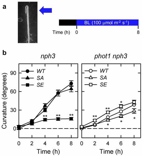
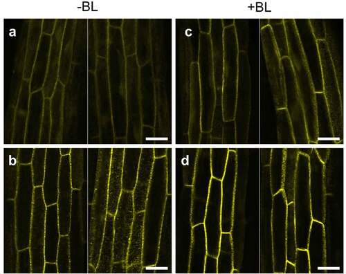
Figure 2. Localization pattern changes for YFP-NPH3 proteins in the phot1 nph3 mutants in response to blue light irradiation. Two-day-old etiolated seedlings of the 35Spro:YFP-NPH3SA phot1 nph3 (a, c) and 35Spro:YFP-NPH3SE phot1 nph3 (b, d) lines were irradiated (c, d: +BL) or not (a, b: -BL) with blue light at 100 µmol m–2 s–1 for 6 h. YFP signals were detected under a TCS-SP5 confocal laser scanning microscope (Leica Microsystems), as described previously.Citation13 Two representative images are shown. White bar, 25 µm.