Figures & data
Figure 1. Study range (in red) with samples collection site indicated by an asterisk.
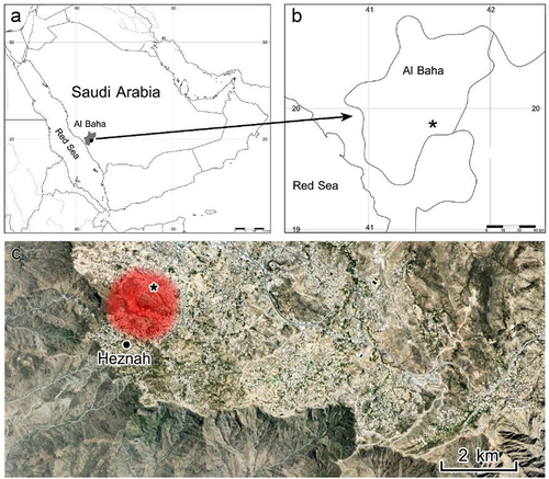
Figure 2. (a-c) C. rosea in microhabitats of the study area. (d) C. rosea with the root system.
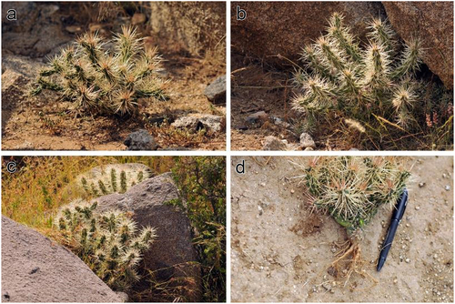
Figure 3. Spine (a, b) and glochid (c, d) micromorphology of C. rosea. Note the barbs that cover the surface of both spines and glochids in SEM images (b-d). tr: trichomes, gl: glochids.
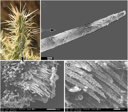
Figure 4. (a, b) surface of the stem with epicuticular waxes covering the epidermis (SEM image in B) and after removal by chloroform (SEM image in C).
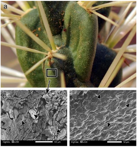
Figure 5. A. Transverse sections (T.Ss.) in the stem revealed the inner structure, with light microscopy images (b-e). B. epidermis with hypodermis and chlorenchyma cells. C. Wide-band tracheids in transverse section (T.S.) and longitudinal section (L.S.) (d). Arrowheads show the primary wall bowing inward between annular rings of the secondary wall. E. pith with druse crystal in some cells. cu: cuticle, dr: druse crystal, ep: epidermis, hyp: hypodermis, pal: palisade cells, t: tracheid, wbt: wide-band tracheids.
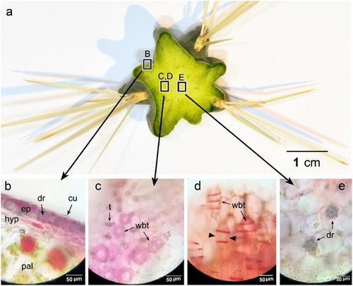
Table 1. Some morpho-anatomical and physiological parameters of C. rosea.
Figure 6. A., C. stem at turgid state. B., D. stem at dry state. Note: high shrinkage of the stem in the dry state.
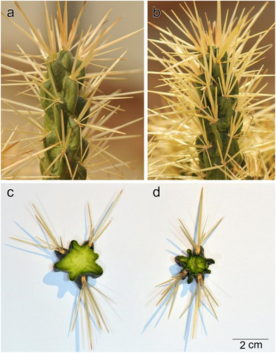
Figure 7. A. inner cortex cells in a turgid state. B. inner cortex cells in a dry state. Note highly undulated cell walls in the dry state (Arrows).
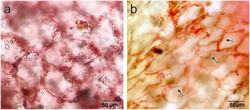
Figure 8. Mature root anatomy. (a) light microscopy image, (b) SEM image. Note: fibrous wood with many vessel elements and large unlignified rays (ray parenchyma) between them containing many druse crystals. (c) SEM image of fibrous wood with vessel elements. (d) SEM image of longitudinal section (L.S.) in a vessel element showing circular bordered pits in wall thickening. pe: periderm, cor: cortex, fw: fibrous wood, ra: ray, dr; druse crystal.
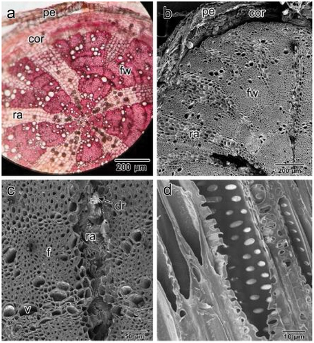
Figure 9. A. druse crystal in ray parenchyma of the root. Note: starch grains on the left. B. EDX spectrum of druse crystal showing a high percentage of Calcium.
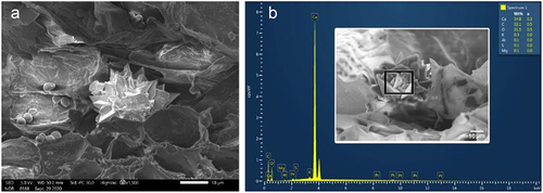
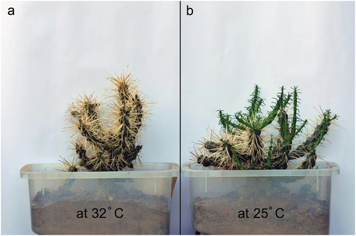
Figure 10. A. C. rosea grown at a mean temperature of 32° in a fully exposed condition (unshaded), and B. grown at a mean temperature of 25° in a partially shady condition. Note new stem segments growth at 25°.