
Figures & data
Figure 1. Optical microscope image of an HBL-100 cell and of a trapped microbead. (a) A microbead (indicated by the white arrow) is first trapped near the HBL-100 cell (cell is in focus) cultured on the coverslip; (b) the cell is displaced vertically and laterally to get the microbead in contact with the cell membrane (microbead is in focus). The scale bar represents 10 µm.
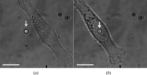
Figure 2. Experimental approach for cell vertical indentation by optical tweezers.
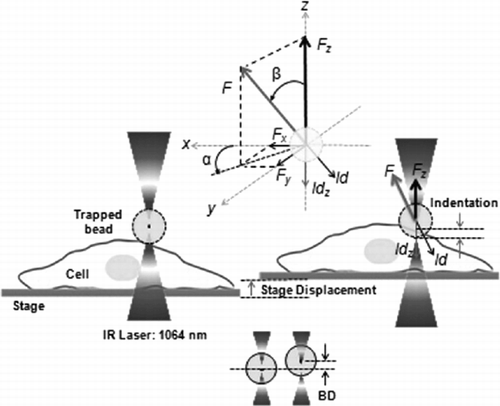
Figure 3. Displacement of the stage (cell) and of the microbead in the trap during the experiment. The sinusoidal movement of the stage make the microbead displacements: in x, y and z. In the first half of the period, the cell moves down and hence the microbead moves freely in the trap. At the beginning of the second half of the period, when the cell intercepts the microbead and pushes it up, the displacement of the bead in z begins to be significant and it is accompanied by smaller lateral displacements in x and y.
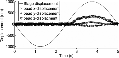
Figure 4. Force excursion vs time during the vertical indentation. (a) Total force F and force components: Fx, Fy and Fz; (b) The orientation of the total force F: angle α and angle β (angles defined in Figure 1).
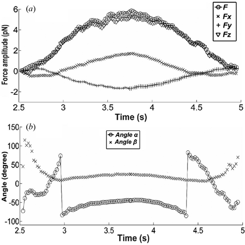
Figure 5. Force-indentation curve for total force F, and the vertical force Fz. The dashed lines define the force range where the fit is applied.
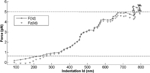
Table 1. Elastic moduli of HBL-100 and MDA-MD-231 cells measured for indentation and retraction, resulting from the total and vertical forces; SD – standard deviation; cell number for each cell line n = 10.
Figure 6. Elastic modulus values calculated for HBL-100 and MDA-MB-231 cells during (a) Indentation and (b) Retraction, using total force (F) and vertical force (Fz). (t-test: ***p < 0.001).
