1,468
Views
14
CrossRef citations to date
0
Altmetric
Articles

Polymer–metal complex nanoparticles-containing cisplatin and amphiphilic block copolymer for anticancer drug delivery

, &
Pages 334-344 | Received 12 Feb 2013, Accepted 23 May 2013, Published online: 11 Oct 2013

Figures & data

Table 1. Molecular weights and compositions of synthesized polymers.

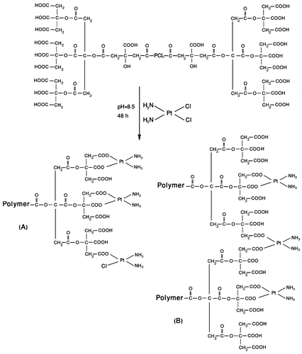
Figure 1 Schematic structure of PCA–PCL–PCA–cisplatin.

Figure 1 Schematic structure of PCA–PCL–PCA–cisplatin.

Figure 2 Particle size distribution of PCA–PCL–PCA–cisplatin nanoparticles at 25 °C in water measured by DLS.
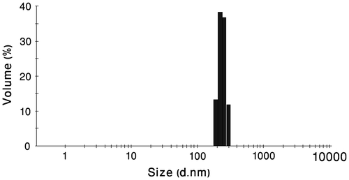
Figure 2 Particle size distribution of PCA–PCL–PCA–cisplatin nanoparticles at 25 °C in water measured by DLS.

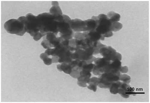
Figure 3 TEM image of PCA–PCL–PCA–cisplatin nanoparticles.

Figure 3 TEM image of PCA–PCL–PCA–cisplatin nanoparticles.

Table 2. Parameters of the prepared nanoparticles.

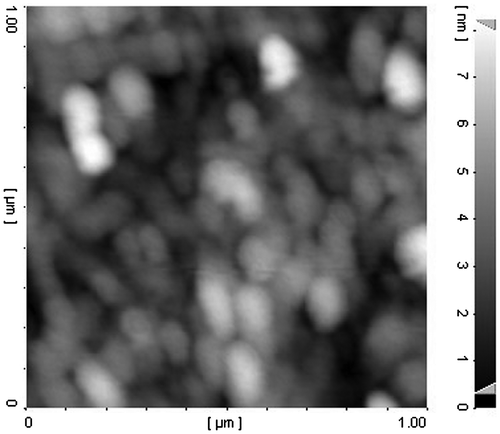
Figure 4 AFM image of PCA–PCL–PCA–cisplatin nanoparticles.

Figure 4 AFM image of PCA–PCL–PCA–cisplatin nanoparticles.

Figure 5 (a) Surface zeta potential of blank PCA–PCL–PCA micelles measured by DLS. (b) Surface zeta potential of PCA–PCL–PCA–cisplatin nanoparticles measured by DLS.
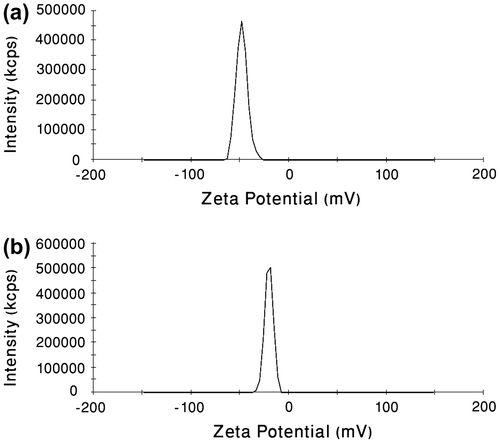
Figure 5 (a) Surface zeta potential of blank PCA–PCL–PCA micelles measured by DLS. (b) Surface zeta potential of PCA–PCL–PCA–cisplatin nanoparticles measured by DLS.

Figure 6 (a) Excitation spectra of pyrene as a function of copolymer concentration at room temperature in water (λem = 393) for PCA–PCL–PCA–cisplatin. (b) Plot of the intensity ratio I336/I333 (from pyrene excitation spectra) vs. log (C) for PCA–PCL–PCA–cisplatin.
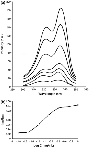
Figure 6 (a) Excitation spectra of pyrene as a function of copolymer concentration at room temperature in water (λem = 393) for PCA–PCL–PCA–cisplatin. (b) Plot of the intensity ratio I336/I333 (from pyrene excitation spectra) vs. log (C) for PCA–PCL–PCA–cisplatin.

Figure 7 Cisplatin release profile from PCA–PCL–PCA–cisplatin nanoparticles in phosphate-buffered saline (PBS, pH 7.4, 0.14 M NaCl) at 37 °C.
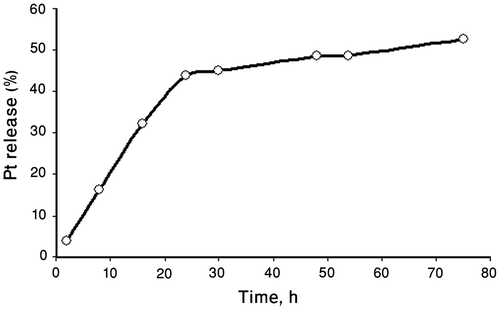
Figure 7 Cisplatin release profile from PCA–PCL–PCA–cisplatin nanoparticles in phosphate-buffered saline (PBS, pH 7.4, 0.14 M NaCl) at 37 °C.

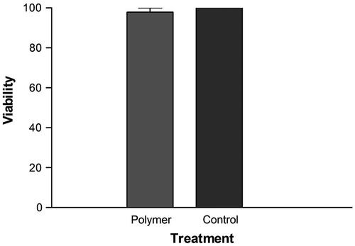
Figure 8 Viability of AE cells treated with blank PCA–PCL–PCA micelle at 1 mg mL−1 for 24 h.

Figure 8 Viability of AE cells treated with blank PCA–PCL–PCA micelle at 1 mg mL−1 for 24 h.

Figure 9 MTT assay to measure the cytotoxicity of PCA–PCL–PCA–cisplatin nanoparticles against HeLa cell line in comparison with cell culture media, blank PCA–PCL–PCA micelle and free cisplatin.
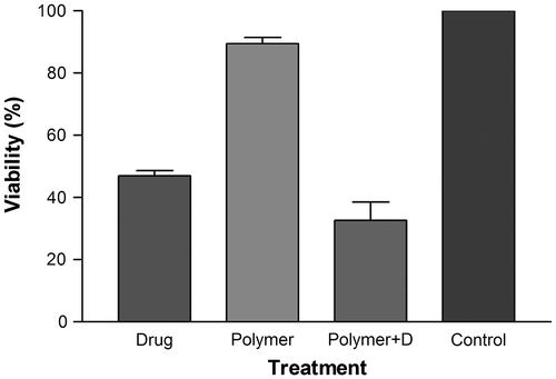
Figure 9 MTT assay to measure the cytotoxicity of PCA–PCL–PCA–cisplatin nanoparticles against HeLa cell line in comparison with cell culture media, blank PCA–PCL–PCA micelle and free cisplatin.
Reprints and Corporate Permissions

Please note: Selecting permissions does not provide access to the full text of the article, please see our help page How do I view content?

To request a reprint or corporate permissions for this article, please click on the relevant link below:

Academic Permissions

Please note: Selecting permissions does not provide access to the full text of the article, please see our help page How do I view content?

Obtain permissions instantly via Rightslink by clicking on the button below:

If you are unable to obtain permissions via Rightslink, please complete and submit this Permissions form. For more information, please visit our Permissions help page.