Figures & data
Figure 1. TEM images of monodispersed silica nanoparticles (bare silica nanoparticles).
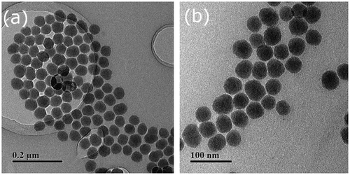
Figure 2. TEM images of hybrid nanoparticles SiO2-g-P(SPMA-co-MAA).
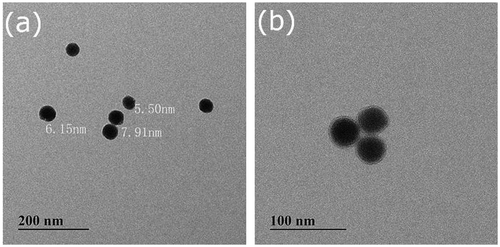
Figure 3. EDS spectrograms of (a) bare silica nanoparticles and (b) hybrid silica nanoparticles coated with P(SPMA-co-MAA).
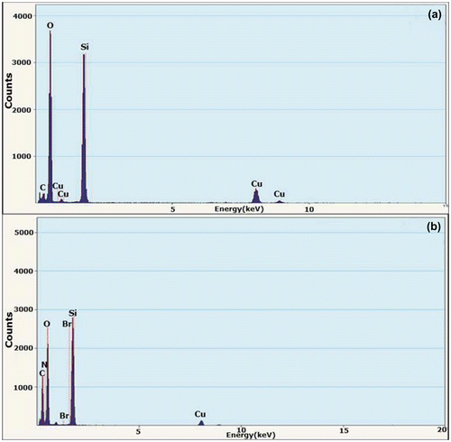
Figure 4. FT-IR spectra of (a) bare silica nanoparticles and (b) hybrid silica nanoparticles coated with P(SPMA-co-MAA).
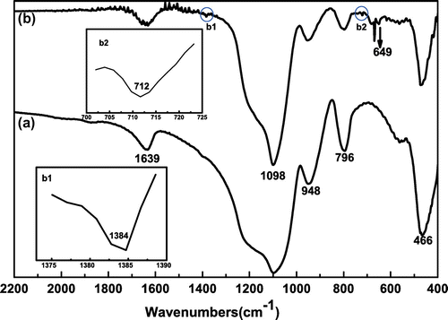
Figure 5. XPS spectra of (a) SiO2, (b) SiO2–Br, and (c) SiO2-g-P(SPMA-co-MAA).
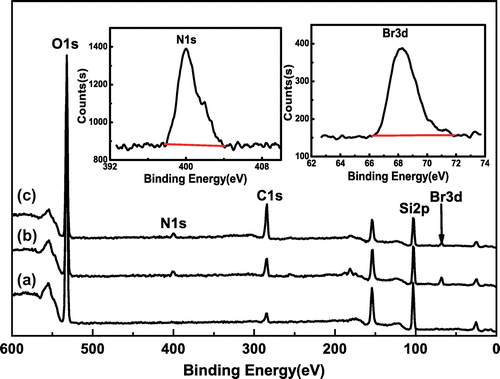
Table 1. Surface elements composition of SiO2, SiO2–Br, and SiO2-P(SPMA-co-MAA).
Figure 6. High-resolution XPS spectra and curve fitting of O1s of (a) SiO2 and (b) SiO2-P(SPMA-co-MAA).
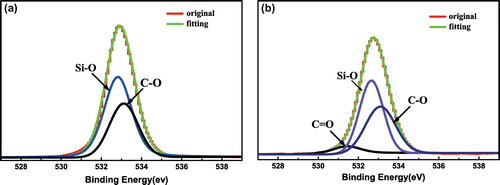
Table 2. O1s high-resolution XPS analysis of SiO2 and SiO2-P(SPMA-co-MAA).
Figure 7. The thermogravimetric analysis of (a) SiO2, (b) SiO2–NH2, (c) SiO2–Br, and (d) SiO2-P(SPMA-co-MAA).
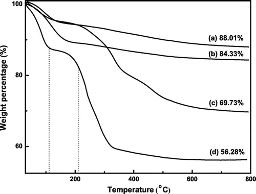
Figure 8. UV–vis spectra of SiO2-g-P(SPMA-co-MAA) in THF under different light conditions.
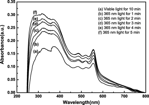
Figure 9. The hydrodynamic diameters distributions of SiO2-g-P(SPMA-co-MAA) under different light conditions.
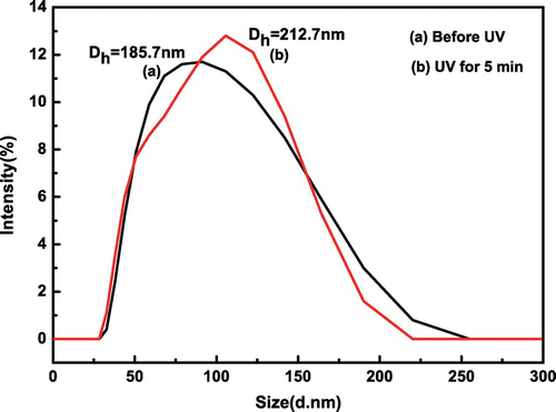
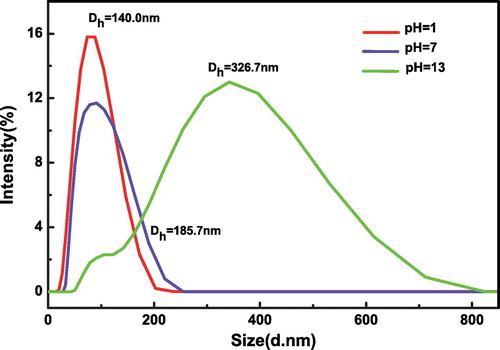
Figure 10. The hydrodynamic diameters distributions of SiO2-g-P(SPMA-co-MAA) in aqueous solutions with different pH values.
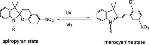
Scheme 1. The photochromism scheme of spiropyrans under UV and vis light.
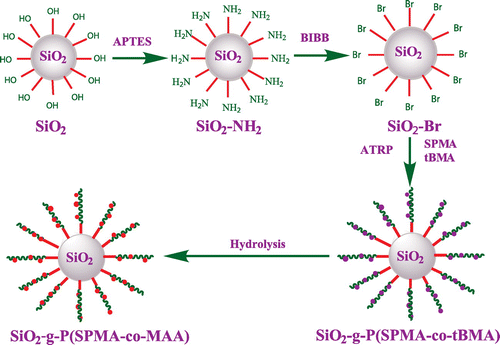
Scheme 2. Synthetic procedure for SiO2-g-P(SPMA-co-MAA).
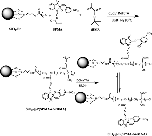
Scheme 3. Synthesis route of SiO2-g-P(SPMA-co-MAA) by SI-ATRP and chemical hydrolysis.
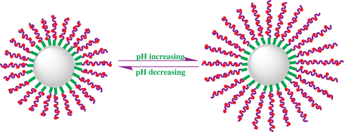
Scheme 4. Schematic illustration of the reversible pH-tunable swelling/shrinking transition.