1,165
Views
4
CrossRef citations to date
0
Altmetric
Articles

Synthesis of G0 aminopolyol and aminosugar dendrimers, controlled by NMR and MALDI TOF mass spectrometry

, , , &
Pages 144-156 | Received 03 Mar 2016, Accepted 29 Aug 2016, Published online: 14 Oct 2016

Figures & data

Table 1. The amounts of reactants and solvents used in the synthesis of dendrimers 1–6.

Figure 1. General reaction scheme.

Figure 1. General reaction scheme.

Figure 2. The structure of the substrates IV, used for the synthesis of dendrimers 16 G0 (I and II – triepoxides, III–V – aminopolyols).

Figure 2. The structure of the substrates I–V, used for the synthesis of dendrimers 1–6 G0 (I and II – triepoxides, III–V – aminopolyols).
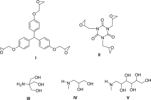
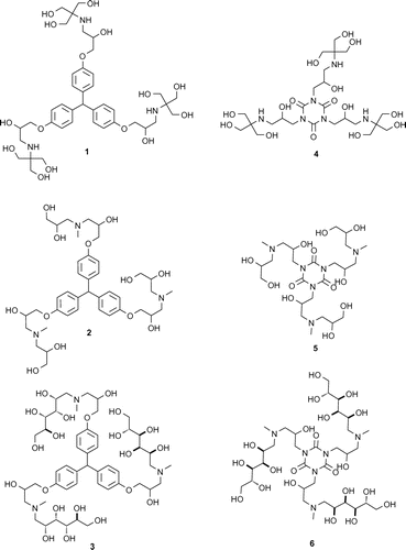
Figure 3. Compounds obtained (16) and used in MS/MS experiment.

Figure 3. Compounds obtained (1–6) and used in MS/MS experiment.

Figure 4. MALDI TOF mass spectrum of: a) a crude compound 1, matrix DHB. Ions: A = [1+H]+ i.e. [I+3Tris+H]+, m/z 824, B = [I+2Tris+H]+, m/z 703; D = [I+DHB+CH3OH+H]+, m/z 647, E =[I+Tris+H2O+H]+., m/z 599; b) purified compound 1, c) crude compound 4.

Figure 4. MALDI TOF mass spectrum of: a) a crude compound 1, matrix DHB. Ions: A = [1+H]+ i.e. [I+3Tris+H]+, m/z 824, B = [I+2Tris+H]+, m/z 703; D = [I+DHB+CH3OH+H]+, m/z 647, E =[I+Tris+H2O+H]+., m/z 599; b) purified compound 1, c) crude compound 4.

Table 2. Main ions (m/z, % r.a.) in MALDI TOF positive mass spectra of crude products 1–6 and proposed ion structures; APO = aminopolyol.

Table 3. Chemical shifts of proton (1–6) and carbon (2, 4–6) atoms δ (ppm), obtained from NMR spectra, measured in D2O; atom labeling given above the table.

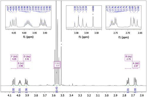
Figure 5. 1H NMR spectrum of compound 4 (D2O, 298 K).

Figure 5. 1H NMR spectrum of compound 4 (D2O, 298 K).

Figure 6. 1H-13C HSQC NMR spectrum of compound 4 (D2O, 298 K) in the range 2.4–4.3 ppm (1H) and 30–180 ppm (13C).
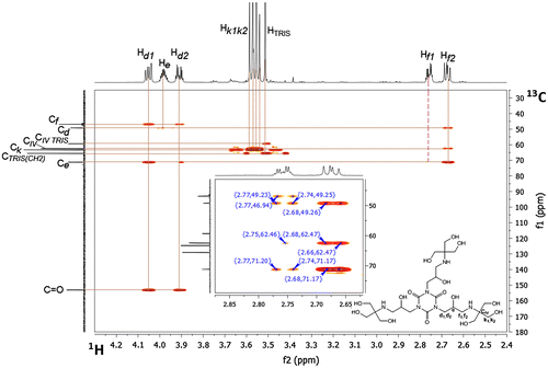
Figure 6. 1H-13C HSQC NMR spectrum of compound 4 (D2O, 298 K) in the range 2.4–4.3 ppm (1H) and 30–180 ppm (13C).

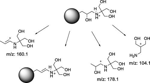
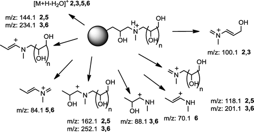
Figure 7. Proposed structure of most intense fragment ions in MALDI MSMS spectra of 16.

Figure 7. Proposed structure of most intense fragment ions in MALDI MSMS spectra of 1–6.

Table 4. Full set of fragment ions, found in MSMS MALDI-QTOF experiment of [M+H]+ ions of compounds 1–6.

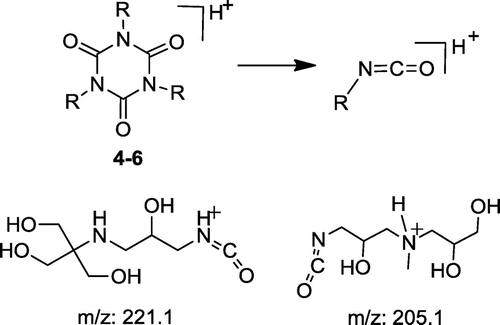
Figure 8. Typical fragmentation for isocyanuric core dendrimers.

Figure 8. Typical fragmentation for isocyanuric core dendrimers.

Figure 9. Common fragmentation pattern of compounds 1 and 4.

Figure 9. Common fragmentation pattern of compounds 1 and 4.

Figure 10. Common fragmentation pattern of compounds 2, 3, 5 and 6 (n=1 or 4).

Figure 10. Common fragmentation pattern of compounds 2, 3, 5 and 6 (n=1 or 4).