Figures & data
Figure 1. Morphologic changes, FISH analysis and RT-PCR of CEP110-FGFR1 fusion in bone marrow cells at the diagnosis. (a/b) Initial bone marrow showed hypercellular with eosinophilia (black arrow) and monocytosis (red arrow); no definite blasts were found in this specimen (WrighteGiemsa, 1000). (c) Nucleotide sequence analysis of the PCR product showing CEP110/FGFR1 fusion transcript with a breakpoint at exon 38 of the CEP110 gene and at exon 9 of the FGFR1 gene.
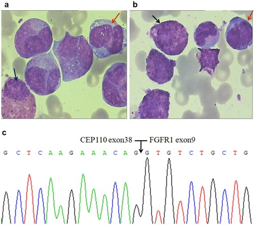
Table 1. Clinical and laboratory features of 16 Cases with CEP110-FGFR1 fusions.
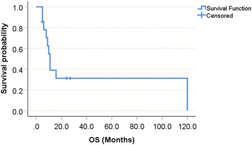
Figure 2. Kaplan–Meier survival analysis of 14 EMS patients with CEP110-FGFR1 fusions (Case 1 and 6 were excluded because of the absence of survival time).