Figures & data
Figure 1. Sequences of gene.1: Proband; 2-5: Proband’s family member; 6: Healthy individual (↓: FGG gene c.1058C>T heterozygous mutation).
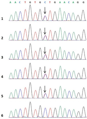
Table 1. Coagulation function of proband and her family members.
Figure 2. Fibrin polymerization curves.
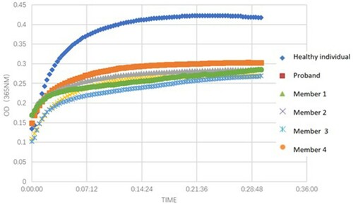
Figure 3. Fibrin clot lysis curves.
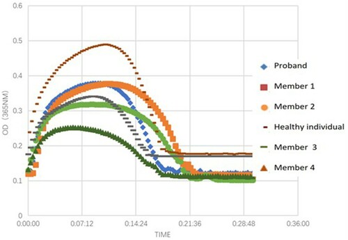
Figure 4. Thromboelastography. 1: Healthy individual; 2-6: Proband and her family members.
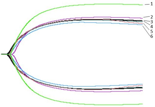
Table 2. Thromboelastography in the proband, her family members, and healthy individual.
Figure 5. Scanning electron microscopy of fibrin clot. A: Healthy individual; B: Proband.
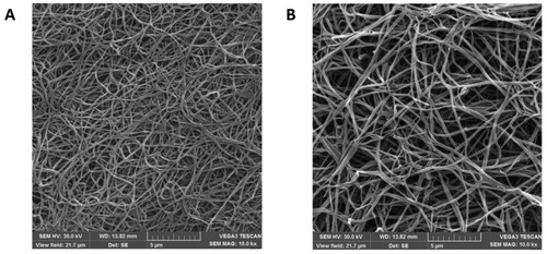
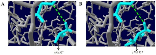
Figure 6. Analysis of the γAla327Val mutation with protein modelling. A: Healthy individual; B: Proband.