Figures & data
Figure 1. Histopathological and immunohistochemical findings. (A) Histiocytes in the background of plasma cells and lymphocytes, exhibiting abundant pale cytoplasm and emperipolesis (arrow). (HE. original magnification ×400); (B, C) Immunoreactive for S-100, CD68, respectively. (original magnification ×200) (D) Negative for CD1a. (original magnification ×200).
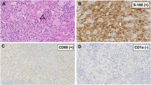
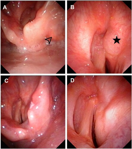
Figure 2. Electronic laryngoscopy of the patient in case 3. (A, B) The pretreatment laryngoscopy revealed lesions located in the right pyriform fossa (arrow) and right anterior ventricular band (star) with mucosal swelling and glottic stenosis. (C, D) After six months of thalidomide treatment, laryngoscopy showed notable remission of lesions in the pyriform fossa and ventricular band.