Figures & data
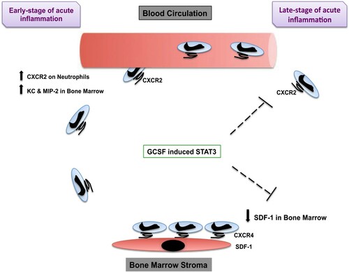
Figure 1. Human G-CSF structure.
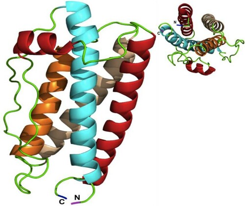
Figure 2. The scheme proposed for domains and downstream signal pathways of G-CSFR.
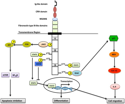
Figure 3. [A] In the absence of the ligand, G-CSFR is associated with Janus kinases (Jaks). [B] The binding of the ligand to the receptor occurs at a 2:2 ligand:receptor subunit stoichiometry, forming a crossover configuration between the receptor subunits that brings the Jaks into proximity and enables their trans-phosphorylation and stimulation. [C] The intracellular 4-tyrosine residues of the G-CSFR (represented by stars) are phosphorylated by Jaks. [D] STAT interacts with the phosphotyrosine residues through their Src Homology 2 (SH2) domains and become phosphorylated by the Jak. Phospho-dimers of STATs accumulate in the nucleus and activate transcription factors that drive the neutrophils from the bone marrow to the blood circulation.
![Figure 3. [A] In the absence of the ligand, G-CSFR is associated with Janus kinases (Jaks). [B] The binding of the ligand to the receptor occurs at a 2:2 ligand:receptor subunit stoichiometry, forming a crossover configuration between the receptor subunits that brings the Jaks into proximity and enables their trans-phosphorylation and stimulation. [C] The intracellular 4-tyrosine residues of the G-CSFR (represented by stars) are phosphorylated by Jaks. [D] STAT interacts with the phosphotyrosine residues through their Src Homology 2 (SH2) domains and become phosphorylated by the Jak. Phospho-dimers of STATs accumulate in the nucleus and activate transcription factors that drive the neutrophils from the bone marrow to the blood circulation.](/cms/asset/7a6bd761-ec42-4c46-9ae8-83b187d0e832/yhem_a_1965725_f0003_oc.jpg)
Figure 4. Scheme of the proposed model. How G-CSF and STAT3 induce the mobilization of neutrophils. At the early stage of acute inflammation, MIP-2, KC, and their shared receptor CXCR2 induce the mobilization of neutrophils from the BM to the blood circulation. At the late stage of acute inflammation, GCSF, together with STAT3, inhibits CXCR2-mediated rapid neutrophil mobilization and reserved neutrophils in the BM, in part throughout their CXCR4 expression, which binds to SDF-1 (stromal cells express SDF-1) expressed in the BM. (↓ reserve ؛ ↑ induce).