Figures & data
Figure 1. Downregulation of miR-204 and upregulation of HGF were observed in AML samples and cell lines. (A–B) The relative expression of miR-204 and HGF mRNA in AML and normal serum samples was analyzed by RT-PCR assay. (C) The relationship between miR-204 and HGF was analyzed. (D) Kaplan-Meier analysis displayed the correlation between miR-204 expression and 5-year overall survival of AML patients. (E) Kaplan-Meier analysis revealed the correlation between HGF expression and 5-year overall survival of AML patients. (F–G) RT-PCR assay was used to detect the expression of miR-204 and HGF in AML cell lines and HS-5 cells. (H) The HGF protein expression was measured by western blot assay. *P < 0.05.
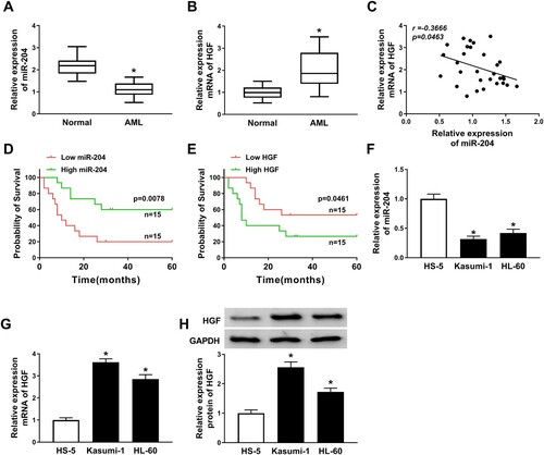
Figure 2. MiR-204 overexpression inhibited AML progression. (A) RT-PCR assay confirmed the efficiency of miR-204 in AML cells. (B–C) Cell proliferation in AML cells transfected with miR-204 or miR-NC was measured by MTT assay. (D and E) Flow cytometry method was employed to detect cell cycle and apoptosis in AML cells transfected with miR-204 or miR-NC. (F–G) Transwell assay was used to determine cell migration and invasion in AML cells transfected with miR-204 or miR-NC. *P < 0.05.
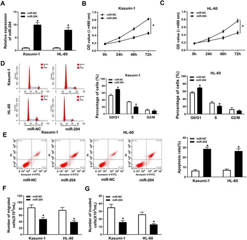
Figure 3. HGF knockdown suppressed AML progression. (A) HGF mRNA expression in Kasumi-1 and HL-60 cells transfected with sh-HGF or sh-NC was detected by RT-PCR. (B-C) HGF protein level in Kasumi-1 and HL-60 cells transfected with sh-HGF or sh-NC was measured by western blot assay and ELISA. (D–E) MTT assay was applied to check cell proliferation in Kasumi-1 and HL-60 cells transfected with sh-HGF or sh-NC. (F–G) The cell cycle and apoptosis of Kasumi-1 and HL-60 cells transfected with sh-HGF or sh-NC were measured by flow cytometry assay. (H–I) Transwell assay was performed to detect cell migration and invasion in AML cells transfected with sh-HGF or sh-NC. *P < 0.05.
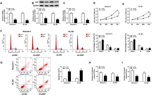
Figure 4. HGF was a target of miR-204. (A)The possible interaction sites between miR-204 and HGF was predicted by MiRcode Tools (http://mircode.org/). (B–E) The target relationship between miR-204 and HGF was confirmed by dual-luciferase assay and RNA pull-down assay. (D–I) The mRNA and protein levels of HGF in AML cells transfected with miR-NC, miR-204, inhibitor miR-204 or inhibitor NC was measured by RT-PCR, western blot assay, and ELISA. *P < 0.05.
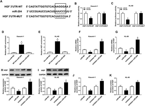
Figure 5. HGF overexpression reversed the effects of miR-204 overexpression on AML cell proliferation, apoptosis, migration, and invasion. (A–B) MTT assay was used to examine cell proliferation in AML cells transfected with miR-NC, miR-204, miR-204+pcDNA-control or miR-204+pcDNA-HGF. (C–D) The cell cycle and apoptosis of AML cells transfected with miR-NC, miR-204, miR-204+pcDNA-control, or miR-204+pcDNA-HGF were assessed by flow cytometry. (F–G) Transwell assay was used to determine cell migration and invasion in AML cells transfected with miR-NC, miR-204, miR-204+pcDNA-control, or miR-204+pcDNA-HGF. *P < 0.05.
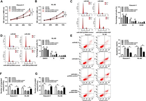
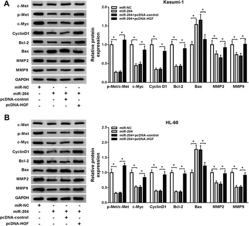
Figure 6. MiR-204 regulated the HGF/c-Met pathway in AML cells. (A-B) The relative protein levels of c-Met, p-Met, c-Myc, Cyclin D1, Bcl-2, Bax, MMP2, and MMP9 in AML cells transfected with miR-NC, miR-204, miR-204+pcDNA-control, or miR-204+pcDNA-HGF were detected by western blot assay. *P < 0.05.