
Figures & data
Figure 1. Sanger sequencing of GPI bα exons in the present case and her parents. (a) The patient had a heterozygous mutation of c.95_101del in chr17:4835994-4836000, father had a heterozygous mutation of c.95_101del, and mother had no mutation. (b) The patient had a heterozygous mutation of c.1012del in chr17:4836911-4836911, father had no mutation, and mother had a heterozygous mutation of c.1012del. (c) The patient had a heterozygous mutation of c.1616T>G in chr17:4837515, father had no mutation, and mother had a heterozygous mutation of c.1616T>G.
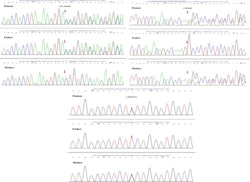
Figure 2. Protein structures of wild-type and mutant GP1BA. (PyMOL 2.5 software was used to generate 3D structural maps of GP1BA wild-type and mutant proteins.): (a) GP1BA wild-type protein, (b) c.95_101del mutant protein, (c) c.1012del mutant protein, (d) part of GP1BA wild-type protein, (e) part of c.1616T>G mutant protein.
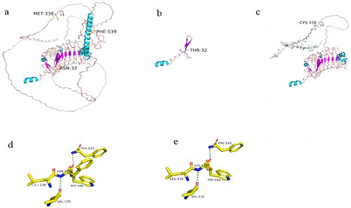
Figure 3. Flow cytometry analysis of GPIbα. (The expression of CD42b on the surface of platelets decreased significantly, while the expression of CD41 and CD61 was normal.)
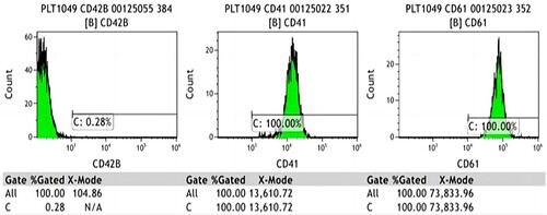
Table 1. Summary of mutation sites reported in previous studies.