Figures & data
Figure 1: Image depicting bilateral ptosis, periorbital oedema and chemosis at initial presentation.

Figure 2: (a) and (b): Coronal and sagittal views of a contrasted brain CT illustrating enlargement and marked contrast enhancement of extraocular muscles (EOM) (inferior (5), medial (1) and superior rectus (3) bilaterally, left lateral rectus (4) and, to a lesser extent, right lateral rectus (4). The superior oblique (2) and optic nerve (6) appeared unaffected.)
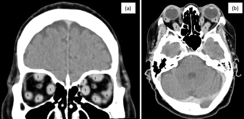
Figure 3: Abdominal CT showing intussusception of the distal ileum (A) with no signs of small bowel obstruction and a desmoplastic reaction (B) with typical findings of linear opacities radiating outwards in a ‘spoke wheel’ or stellate pattern, with ‘in-drawing’ of the surrounding tissues.
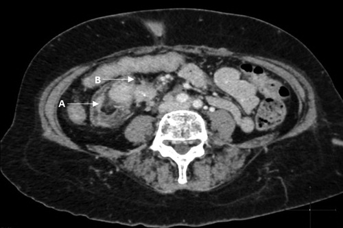
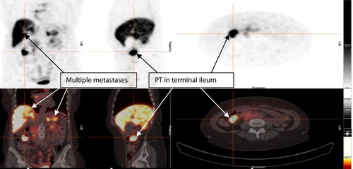
Figure 4: Ga 68 DOTANOC PET showing metastatic lesions and intense uptake of the primary lesion in the terminal ileum, with multiple metastases to liver, spleen and pancreas.