Figures & data
Table 1. Composition of diets (g/kg diet) fed to rats.
Figure 1. Chromatography (HPLC) of mesozeaxanthin.
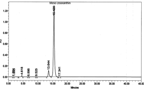
Table 2. Effect of MZ supplementation on the body weight, visceral fat and liver weight in rats fed HFD for 12 weeks.
Table 3. Effects of MZ on biochemical parameter levels in rats fed HFD for 12 weeks.
Table 4. Effects of MZ on the antioxidant status rats fed HFD for 12 weeks.
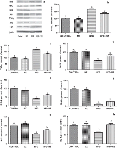
Figure 2. Hepatic NF-κB, TNF-α, BCO2, IRS-1,PPAR-γ Nrf-2 and HO-1 expression levels in mesozeaxanthin (MZ) supplemented high-fat diet (HFD) fed rats and control groups.
Notes: The Western blot strips of the proteins measured in this study are shown in Panel A. Panels B–F show the expression level of NF-κB, TNF-α, BCO2, IRS-1, PPAR-γ, Nrf-2 and HO-1 in various groups. The intensity of the bands shown in Panel A was quantified by densitometric analysis. Data are expressed as a ratio of normal control value (set to 100%). Each bar represents the mean and standard error of mean. Blots were repeated at least three times (n = 3) and only a representative blot is shown in Panel A. β-Actin was included to ensure equal protein loading.