Figures & data
Table 1. Percentage of normal weight and overweight individuals belonging to each sweet taste sensitivity group.
Table 2. Comparison of salivary parameters (mean ± standard error) between sweet taste sensitivity groups.
Table 3. MS identification of the salivary proteins differentially expressed among taste sensitivity groups.
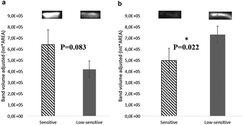
Figure 1. Protein bands differing between the two sweet sensitivity groups.
* Differences are statistically significant for P < 0.05.
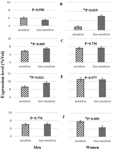
Table 4. Details of mass spectrometry results for protein spots associated with sweet taste sensitivity. Comparisons between sweet taste sensitivity groups are presented.
Figure 2. Representative 2-DE profile of saliva analysed. Circles represent the spots differentially expressed between sensitive and low-sensitive groups.
MW – molecular weight (kDa); pI – isoelectric point.
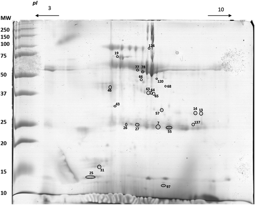
Figure 3. Enzymatic activity of salivary α-amylase (U/min) in men (N = 46) and women (N = 41) with different sensitivity levels to sweet taste (mean ± SEM).
*Statistically significant differences: P < 0.05.
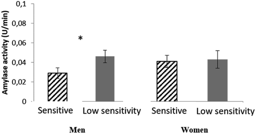
Figure 4. Representative Western blot analysis of cystatins S-SA-SN (a) and CA VI (b) in mixed saliva samples of individuals with different sensitivity levels to sweet taste (mean ± SEM).
*Statistically significant differences: P < 0.05.