Figures & data
Figure 1. MoMic digital microscope scanner (1) with external motor unit attached (2). The microscope glass (3) is placed in the slide holder (4), which is placed in the microscope and navigated from the motor unit. The device is connected to and operated from a laptop computer (5) running software (6) for operation of the device.
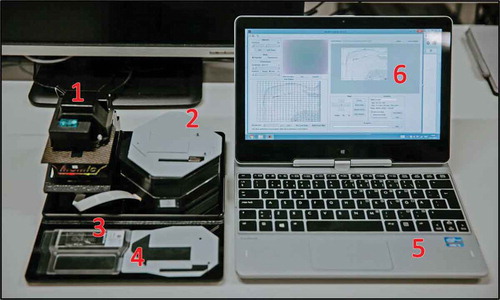
Figure 2. Region of glass slide captured with the mobile microscope. Enlarged areas (right) showing A. lumbricoides eggs in sample (magnified images showing native, full resolution of captured images at 100% digital magnification).
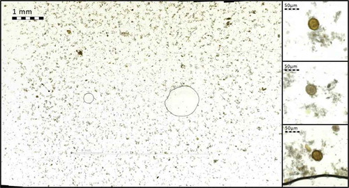
Figure 3. Stool sample showing A. lumbricoides eggs, digitized with (a) mobile microscope, and (b) reference slide-scanner.
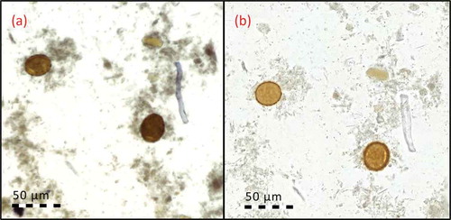
Figure 4. Captured images with the mobile microscopes with visible parasites. Enlarged areas showing the parasites at 300% digital zoom. (a) A. lumbricoides, (b) T. trichiura, (c) hookworm, (d) S. haematobium.
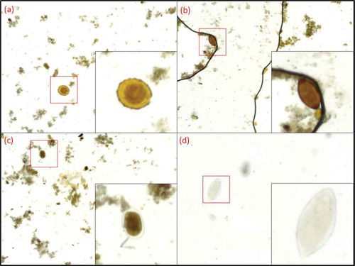
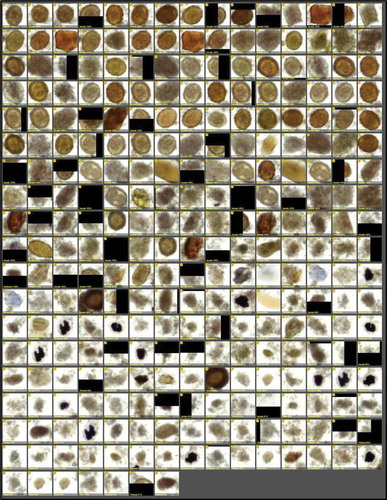
Figure 5. Results of digital image analysis of fixated stool sample (A. lumbricoides infection), showing detected regions of interest of fixated stool sample, sorted by certainty of representing parasite (descending order, from most likely candidates to least likely), as detected by the image analysis software.