Figures & data
Table 1. Demographic and clinical characteristic of women (n = 200).
Figure 1. A healthy skin Phyla that was identify by Vitek 2. Actinobacteria (17%), Firmicutes (79.5%) and Proteobacteria (3.5%).
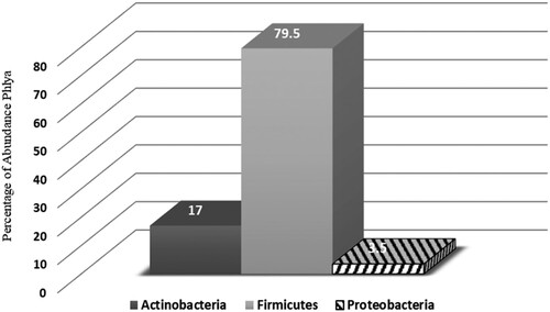
Table 2. Taxonomic identification of skin microbiota of Saudi Females. Relative abundances of the top three phyla across 200 samples included in this study with relative abundance of the top genera within sample.
Table 3. Classification and distributing skin microbiota and its connected microenvironments; moist (Underarm), sebaceous (forehead and behind- ears) and dry (forearm).
Figure 2. Relative abundance of staphylococcal species in current samples. Pie chart performs the abundance of species as a percentage of the entire microbial community.
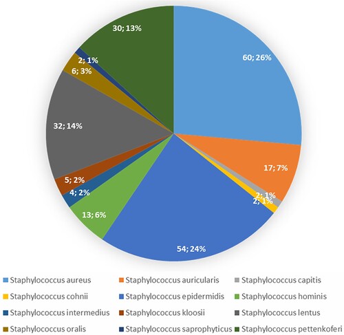
Figure 3. The percentage of Staph. Abundance in different body parts. A significant difference in S. aureus and S. epidermidis distributed between various body parts.
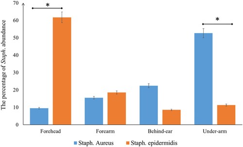
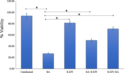
Figure 4. S. epidermidis protects NHEK from the effects of S. aureus. NHEK viability was unaffected by S. epidermidis (S.EPI). S. epidermidis protected the NHEK from the effects of S. aureus (SA) and the viability of NHEK treated with combination of two bacteria was 50.38%. The viability of NHEK treated with S. epidermidis 2 hours before adding the S. aureus was 70.91% compared to 26.87% in NHEK infected with S. aureus alone (P = 0.03, n = 3).