Figures & data
Figure 1. The configuration (left) and the geometries (right) of objects 𝒪1 (up) and 𝒪2 (down).
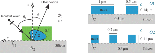
Figure 2. Modulus (up) and phase (down) of the computed (red, dashed line) and measured (black, full line) scattered fields for object 𝒪1 illuminated from direction θ1 = −17.52° (left) and for object 𝒪2 illuminated from direction θ1 = −14.82° (right).
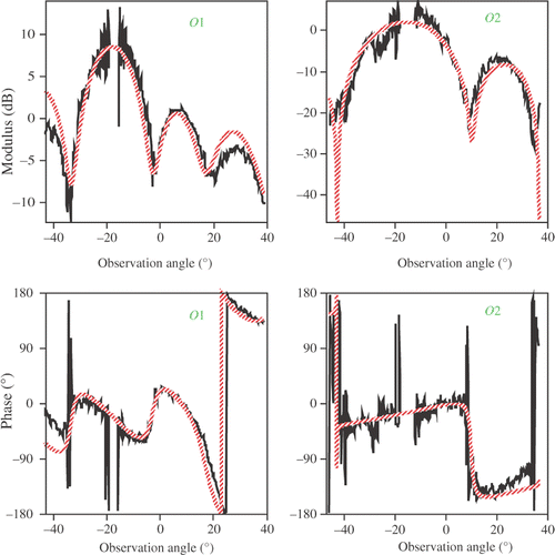
Figure 3. Evolution of the means mk and variances of the contrast for the classes k = 1 (resin) and k = 2 (air) during the iterative process for objects 𝒪1 (left) and 𝒪2 (right): m1 (red), m2 (×50, black),
(×50, green dotted line) and
(×50, blue dashed line).

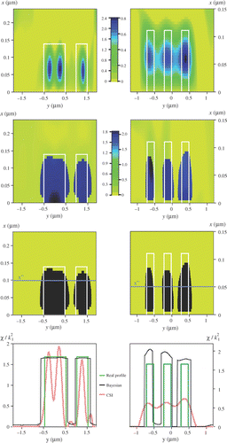
Figure 4. The results obtained for objects 𝒪1 (left column) and 𝒪2 (right column) of : the normalized contrast () retrieved by means of the CSI method (1st row) and by means of the proposed Bayesian approach (2nd row), the class (3rd row) and the normalized contrast profile retrieved at a height x″ (x″ = 0.1 µm for 𝒪1 and x″ = 0.05 µm for 𝒪2) (last row). The true profiles are indicated by white or green lines.