Figures & data
Table 1. Number of iterations needed for the convergence with different starting points for interior and exterior surfaces of the cornea.
Table 2. Results of the numerical experiment for finding true values of parameters and
. The model was constructed from (Equation51
51
51 ) with additive noise on level
and
,
.
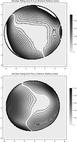
Figure 1. Contur plots of the absolute errors in fitting defined in (Equation53
53
53 ). On the top: exterior surface, on the bottom: interior surface of cornea. Horizontal and vertical axes, shown in millimetres, depict
and
coordinates of the point on the corneal surface. The light colour outside the cornea represents the reference surface.