5,109
Views
1
CrossRef citations to date
0
Altmetric
Review

Inhaled antifungal therapy: benefits, challenges, and clinical applications

Pages 755-769 | Received 23 Dec 2021, Accepted 27 May 2022, Published online: 02 Jun 2022

Figures & data

Figure 1. Schematic illustration of the fate of inhaled Aspergillus conidia, and opportunity of inhaled antifungal therapy. Inhaled conidia or germinated hypha are often cleared up by resident macrophages or neutrophils. Survived hypha is branching and grows, forming biofilm. On some occasions, fungus forms fungal ball in lung lumen, and in immunocompromised condition, fungus invades systemically. There are different treatment options/regimen at different infection stage.

Figure 1. Schematic illustration of the fate of inhaled Aspergillus conidia, and opportunity of inhaled antifungal therapy. Inhaled conidia or germinated hypha are often cleared up by resident macrophages or neutrophils. Survived hypha is branching and grows, forming biofilm. On some occasions, fungus forms fungal ball in lung lumen, and in immunocompromised condition, fungus invades systemically. There are different treatment options/regimen at different infection stage.

Table 1. Preclinical evidence of beneficial effects of inhaled antifungals.

Table 2. Clinical evidence of beneficial effects of inhaled antifungals by off-label use.

Table 3. Novel inhaled antifungals in clinical phase.
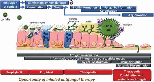
Figure 2. Preclinical in vitro assay method for inhaled antifungals. a) air-liquid interface cultured pseudostratified epithelium (nasal, bronchial, small airway). b) bilayer alveolus model [Citation98]. Black freeform scribble: fungus body. c) A. fumigatus infection in bilayer (apical phase). Histology of alveolus model d) non-infected, e) 48 h post infection with A. fumigatus conidia. Solid arrow: Conidial head and conidiophore, Dotted arrow: hyphae and foot cell, White arrow: original endothelial and epithelial cell bilayer.

Figure 2. Preclinical in vitro assay method for inhaled antifungals. a) air-liquid interface cultured pseudostratified epithelium (nasal, bronchial, small airway). b) bilayer alveolus model [Citation98]. Black freeform scribble: fungus body. c) A. fumigatus infection in bilayer (apical phase). Histology of alveolus model d) non-infected, e) 48 h post infection with A. fumigatus conidia. Solid arrow: Conidial head and conidiophore, Dotted arrow: hyphae and foot cell, White arrow: original endothelial and epithelial cell bilayer.

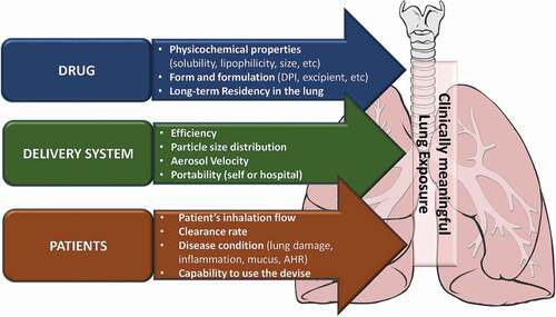
Figure 3. Factors affecting lung exposure and distribution of inhaled-antifungal agent. AHR: airway hyperresponsiveness.

Figure 3. Factors affecting lung exposure and distribution of inhaled-antifungal agent. AHR: airway hyperresponsiveness.