Figures & data
Figure 1. Zipper closure. Small or oval elongated lesions are clipped in a zipper fashion (30), starting in one corner and working clip by clip to the other corner. With every clip that is placed, the edges close to the clip are pulled a bit closer to each other, so the subsequent clips will easier reach the opposite wound margins.
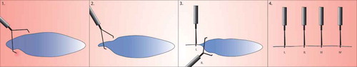
Figure 2. Mechanism of action of TTS clip and Ovesco clip. (a). Endoscopic clipping of the mucosal layer with regular clips. (b). 3. The target tissue is sucked in the cap. The Twingrasper or Anchor devices (Ovesco Endoscopy AG, Tübingen, Germany) can be used to grab the target tissue and pull the margins into the cap, especially in case of tissue induration. 4–5. When the Ovesco clip is deployed, the clip teeth may slightly push the tissue out of the cap. If the margins of the lesion are not completely inside the cap, this may result in incomplete closure.
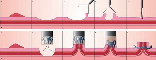
Figure 3. Approximating the peripheral wound edges prior to Zipper closure. Larger or round lesions, where the edges are further apart, can benefit from placing a clip in two opposite peripheries of the wound surface, making it more almond-shaped. After doing this, clipping can be continued between the clips in a zipper fashion.
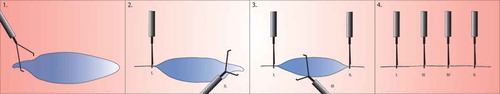
Table 1. Overview of studies on prophylactic clipping.
Table 2. Clinical trials on prophylactic clipping after EMR.
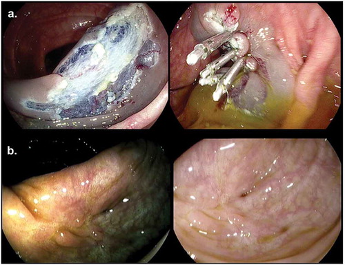
Figure 4. (a). Prophylactic clip closure of resection field after piecemeal EMR. (b). Clip artifact in scar after EMR with HDWLE and Iscan.