
Figures & data
Figure 1. Diagram of Tenckhoff catheter placed in the abdominal cavity for peritoneal dialysis showing placement of the tip in the pelvis and transcutaneous segment. Used with permission from Kidney International (Publisher: Elsevier).
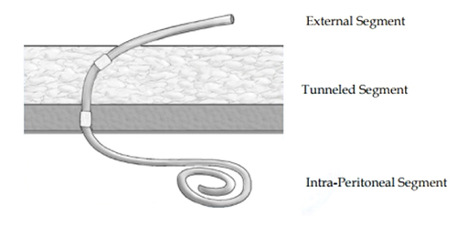
Figure 2. Diagram illustrating the technique for exchange of fluid during peritoneal dialysis. Used with permission from National Institute of Diabetes and Digestive and Kidney Diseases, National Institutes of Health.
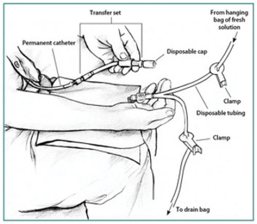
Table 1. Proposed criteria for the diagnosis of exit site infections (reference 22).