Figures & data
Figure 1. Characterization of 25 nm green fluorescent polystyrene nanoparticles (PSNP) in Elendt M4 medium. (a) TEM image after 1 h in solution. Scale bar: 200 nm. (b) size distribution of the hydrodynamic diameter measured by DLS, (c) zeta potential (n = 3), (d) settling of PSNP in medium containing algae over time (colored lines) and background of medium containing algae only (gray lines) measured by a fluorometer, expressed as mg L−1 as approximated by a linear calibration (Figure S1). (For interpretation of the references to color in this figure legend, the reader is referred to the web version of this article.)
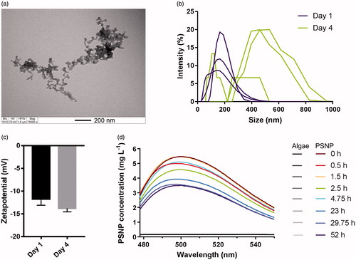
Figure 2. Representative image of PSNP in the intestine and lipid droplets of an adult Daphnia magna exposed to PSNP for 2 days (n = 8). (a) The PSNPs (green) are present inside of the gut but did not cross the epithelial border structurally. The intestinal epithelium is between the basement membrane (dotted line) and the apical membrane (dashed line). Fluorescent spots outside of the gut (arrows) are likely a result of sample preparation (crushing). (b) Accumulation of PSNP in lipid droplets (arrows) of adult animals was not detected using a GFP filter exposure duration of 2.5 s, gain of 2.0 and gamma of 0.6. The exposure duration may be too low to detect PSNP in maternal lipid droplets, however a short exposure duration was necessary to avoid autofluorescence. (For interpretation of the references to color in this figure legend, the reader is referred to the web version of this article.)
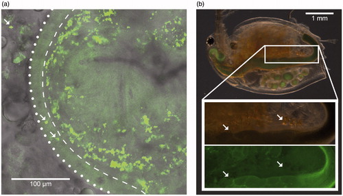
Figure 3. Representative fluorescence microscopy images of different stages of Daphnia magna development (n = 8). Adult daphnids were exposed to 5 mg L−1 PSNP from the moment that their brood pouch was empty until their embryos were in stage 4. GFP filter settings were an exposure duration of 2.5 s, a gain of 2.0 and a gamma of 0.6. Stage 1: the adult daphnid took up PSNPs but the embryo has not. Stage 3: the developing embryos took up PSNPs. Stage 4: PSNPs are concentrated in droplets in the embryo. Stage 6: neonate that has stored PSNPs in its fat droplets.
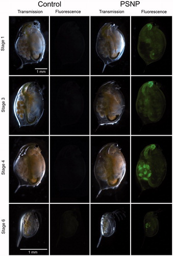
Figure 4. Representative fluorescence microscopy images of neonate Daphnia magna. The yellowish droplets in the central part of the body are fat droplets, as confirmed by staining animals with the lipophilic dye Nile red (bottom picture). DsRED filter exposure settings were an exposure duration of 448.9 ms, a gain of 2.0 and a gamma of 0.6.
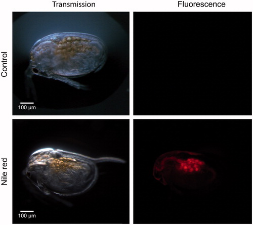
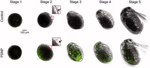
Figure 5. Representative confocal fluorescence microscopy images of Daphnia magna embryos during ex vivo exposure (n = 24). Overlay images of bright field and excitation at a wavelength of 488 nm are depicted of stage 1 to 5 showing uptake of PSNP (green fluorescent) from stage 2 onwards. Embryos at stage 2 have an inner (white arrow) and outer (black arrow) chorion, the latter bursts before stage 3. (For interpretation of the references to color in this figure legend, the reader is referred to the web version of this article.)