
Figures & data
Figure 1. Overview of pre-conceptional exposure of female mice, mating as well as postnatal OVA exposure of offspring. (A) Mucosal tolerance was induced by weekly i.n. instillations of 5 µg OVA in offspring aged 4-8 weeks. This was followed by allergen immunization at age 9 weeks (5 µg OVA in 1 mg Al(OH)3) and a booster of 5 µg OVA one week later. Finally, offspring were exposed to a 0.001% OVA aerosol by inhalation. (B) Suboptimal sensitization (5 µg OVA in 1 mg Al(OH)3, no booster) and inhalation of allergen (1% OVA) by was performed in offspring, starting at 2 weeks of age.
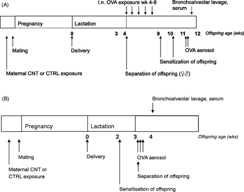
Figure 2. Effect of pre-conceptional maternal CNT exposure on mucosal tolerance in offspring. After 5 weeks of i.n. OVA exposure, offspring were s.c. immunized with OVA and exposed to OVA by inhalation, c.f. . OVA-specific IgE (A) and IgG1 (B) were measured in serum and the total number of cells was counted in recovered BALF (C). Individual and median values of female (black) and male (white) offspring are shown (n = 7-8). p-values of the GLM analyses are given above B and C. Broken line indicates the lower detection limit of the ELISA.
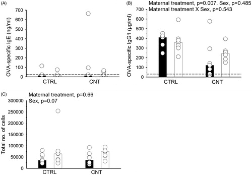
Figure 3. Effect of pre-conceptional maternal CNT exposure on allergen immunization in offspring. At age 2 weeks, offspring were s.c. immunized with OVA and subsequently exposed to OVA by inhalation, c.f. . OVA-specific IgE (A) and IgG1 (B) were measured in serum and the total number of spleen cells counted (C). Individual and median values of female (black) and male (white) offspring are shown (n = 7, except for CTRL offspring, where n = 5 for the IgG1 measurements). p-values of the GLM analyses are given above the figures. Data in (B) were log-transformed for the statistical analysis. Broken line indicates the lower detection limit of the ELISA.
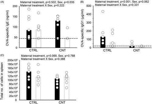
Figure 4. Effect of pre-conceptional maternal CNT exposure on allergen immunization in offspring. At age 2 weeks, offspring were s.c. immunized with OVA and exposed to OVA by inhalation, c.f. . Allergic airway inflammation was assessed in BALF from the total number of cells (A), the percentage (B) and total number (C) of eosinophils and from protein levels (D). Individual and median values of female (black) and male (white) offspring are shown (n = 7). p-values of the GLM analyses are given above the figures. Data in (A) and (C) were log-transformed for the statistical analyses.
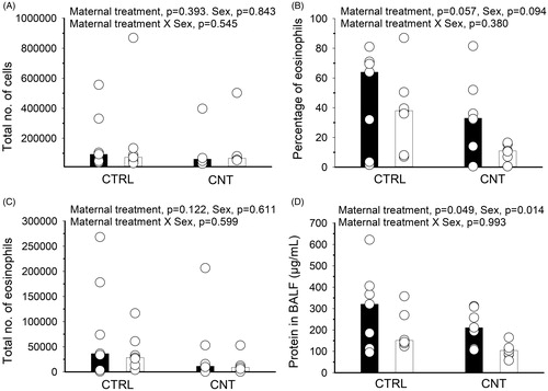
Figure 5. Effect of pre-conceptional maternal CNT exposure on IgM levels in naïve 4-week-old offspring (A, n = 4-5 per group per experiment) and suboptimally sensitized offspring (B, n = 4-5, from ). In B, individual and median values of female (black) and male (white) offspring are shown. The limit of detection for the assay was 3990 ng/mL. p-values of the GLM analyses are given above the figures. All data were log-transformed for the statistical analyses.
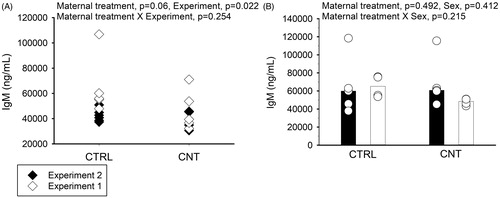
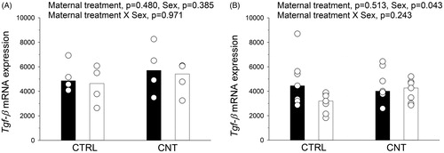
Figure 6. Tgf-β mRNA expression was quantified in (A) spleens from naïve offspring at postnatal day 7 (n = 4), and in (B) lung tissue from the sensitized offspring (n = 7, from ). Individual and median values of female (black) and male (white) offspring are shown. p-values of the GLM analyses are given above the figures.
Supplemental Material
Download PDF (464.7 KB)Data availability statement
The data that support the findings of this study are available from JSH and KSH, upon reasonable request.