Figures & data
Figure 1. Map of of four ECOMAR target sites.
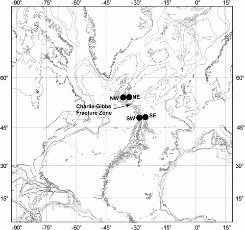
Table I. Data on stations of ECOMAR cruises
Figure 2. Hansenothuria sp. (A–C) Underwater photographs: (A) St. JC048/43 Dive 174; (B) St. JC048/40 Dive 173; (C) JC048/53 Dive 178. (D–G) Ossicles of papillae. (D) scale 0.1 mm; (E–G) scale 0.2 mm.
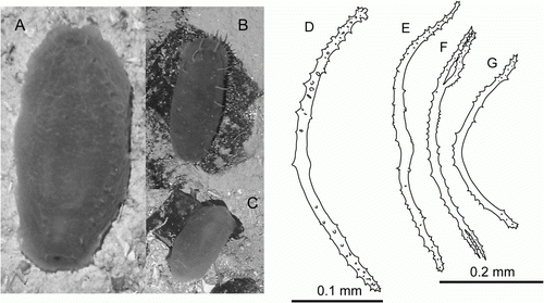
Figure 3. Synallactes aff. crucifera, St. 48/16 Dive 162. (A–C) preserved specimen in dorsal, ventral, and lateral view; (D,E) dorsal ossicles, upper and side view; (F,G) ventral ossicles, upper and side view.
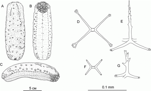
Figure 4. Synallactidae gen. et sp. indet. (A,B) Underwater photographs: (A) St. JC048/40 Dive 173; (B) St. JC048/43 Dive 174. (C–E) St. JC048/43 Dive 174, ossicles of papillae, scale 0.05 mm; (F) St. JC048/43 Dive 174, tentacle ossicles, scale 0.2 mm; (G) St. JC048/43 Dive 174, gonad ossicles, scale 0.2 mm.
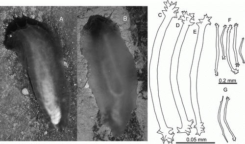
Figure 5. Laetmogone billetti sp. nov., paratype, St. JC048/16, dive 162. (A,B) Dorsal and ventral view, scale 2 cm; (C) tentacle disc magnified; (D–H) dorsal ossicles, scale 0.1 mm; (I,J) ventral ossicles, scale 0.1 mm.
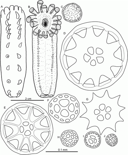
Figure 6. Laetmogone billetti sp. nov., paratype, St. JC048/16, dive 162. (A–G) Tube feet; (H–L) papillae; (M–V) tentacles; (U) magnified ossicle (T). (F,U) scale A; all others scale B.
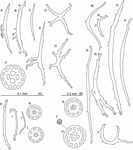
Figure 7. (A–E) Amperima furcata (Hérouard, Citation1899). (A,B) St. JC048/43 Dive 174, dorsal and ventral view, scale A; (C–E) dorsal ossicles, St. JC048/54, Dive 179: (C) scale B; (D,E) scale C. (F–N) Ellipinion delagei (Hérouard, Citation1896), St. JC048/24 Dive 165. (F,G) Dorsal and ventral view, scale D; (H) distal part of tube foot, magnified; (I) ventral C-shaped ossicle, scale E; (J,K) ventral rods, scale F; (L–N) tube foot ossicles, scale G.
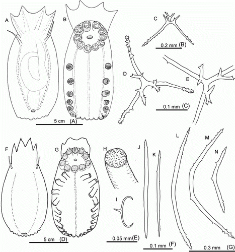
Figure 8. Ellipinion alani sp. nov., holotype. (A) Dorsal view, scale A; (B–F) ventral ossicles; (G–M) tube foot ossicles; (N–R) tentacle ossicles. (D–F,M) scale B; (B,C,G–L,N–R) scale C.
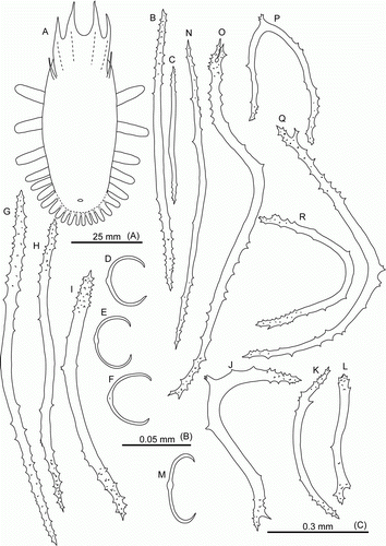
Figure 9. Peniagone azorica von Marenzeller, 1893, dorsal ossicles, side view, ha. (A–D) St. JC048/24 Dive 165; (E–G) St. JC011/101; (H–L) BIOICE Sample No. 2852. (da) Deflection angle.
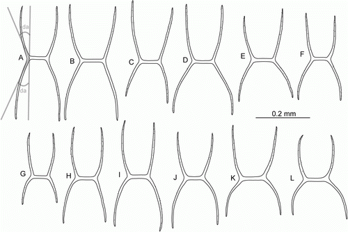
Figure 10. Peniagone islandica Deichmann, Citation1930, dorsal ossicles, side view. (A–D) St. JC048/43 Dive 174; (E–G) St. JC037/15; (H,I) St. JC048/54 Dive 179.
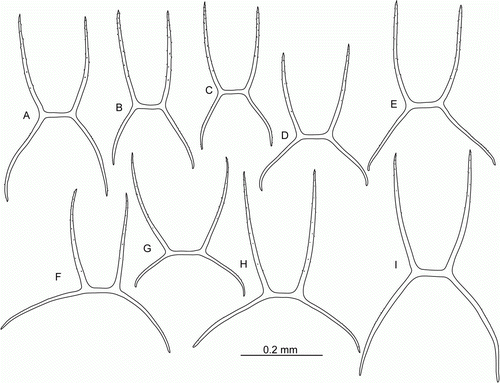
Figure 11. Peniagone islandica Deichmann, Citation1930, dorsal ossicles, side view. (A–E) St. JC048/53 Dive 178; (F–J,K–M) two specimens from St. JC037/15.
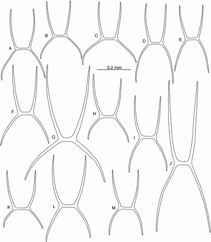
Figure 12. (A–G) Peniagone islandica Deichmann, Citation1930, St. JC048/53 Dive 178. (A,B) Dorsal and ventral view, scale A; (C–E) dorsal ossicles, scale C; (F,G) ventral ossicles, scale B. (H–J) Peniagone azorica von Marenzeller, 1893, St. JC011/101. (H,I) Dorsal ossicles, scale C; (J) ventral ossicles, scale B. (K–M) Peniagone longipapillata Gebruk, Citation2008. (K) Dorsal view, St. JC048/16 Dive 162, scale A; (L) dorsal ossicle of Peniagone-type, St. JC048/24 Dive 165, scale D; (M) dorsal rods, St. JC048/24 Dive 165, scale E.
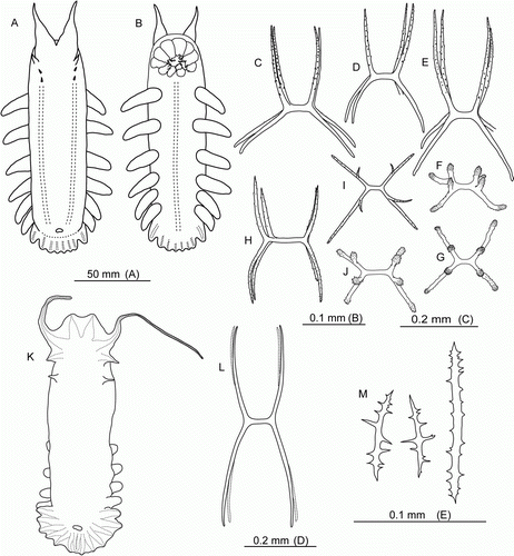
Figure 13. Peniagone coccinea sp. nov. (A) Paratype, St. JC048/43 Dive 174, dorsal view, scale 50 mm; (B,C) St. JC037/15, freshly caught specimen, scale 50 mm; (D–I) dorsal ossicles, scale 0.1 mm; (J–L) ventral ossicles scale 0.1 mm: (E,G,H,I,L) holotype; (D,F,J,K) St. JC037/15.
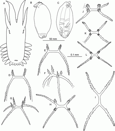
Figure 14. Peniagone coccinea sp. nov., St. JC048/43 Dive 174. (A–G) Tube foot ossicles; (H–P) tentacle ossicles.
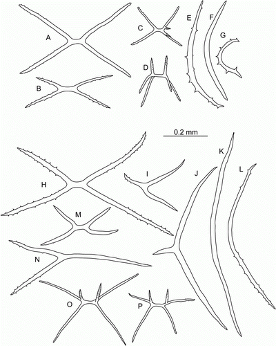
Figure 15. Molpadia aff. blakei (Théel, 1886), St. JC037/27. (A–H) Body wall ossicles; (I–L) tail ossicles.
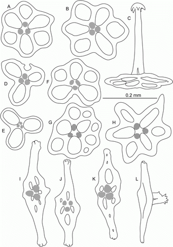
Figure 16. (A,B) Labidoplax sp., St. JC037/15, body wall ossicles. (C,D) Gephyrothuria alcocki Koehler & Vaney, Citation1905. St. JC037/19. (C) Dorsal view, scale 25 mm; (D) tentacle disc, magnified. ap, anal papilla; gp, genital papilla; eg, endgut; pp, posterior protuberance.
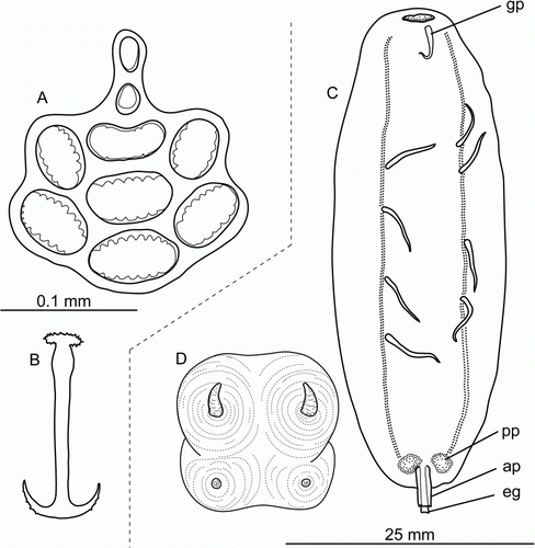
Figure 17. Underwater images. (A) Staurocucumis abyssorum, St. JC048/43 Dive 174; (B) Benthothuria funebris, St. JC048/29 Dive 169; (C) Hansenothuria sp., St. JC048/40 Dive 173; (D) Mesothuria maroccana, St. JC048/40 Dive 173; (E) Benthodytes gosarsi, St. JC048/3 Dive 158; (F) Psychropotes depressa, St. JC048/43 Dive 174; (G) Psychropotes depressa, St. JC048/54 Dive 179; (H) Laetmogone billetti sp. nov., St. JC048/24 Dive 165; (I) Laetmogone billetti sp. nov., St. JC048/16 Dive 162; (J) Peniagone azorica, St. JC048/24 Dive 165; (K) Amperima furcata, St. JC048/43 Dive 174; (L) Myriotrochus clarki, St. JC048/24 Dive 165; (M) Peniagone islandica and Amperima furcata, St. JC048/43 Dive 174; (N) Ellipinion alani sp. nov., St. JC048/24 Dive 165; (O) Peniagone longipapillata, St. JC048/24 Dive 165.
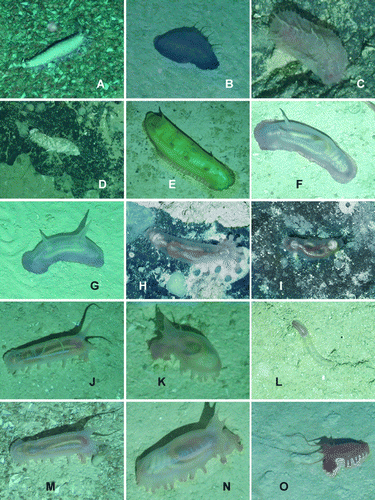
Figure 18. (A–J) Underwater images; (K–P) freshly caught specimens from ROV, photos courtesy: David Shale. (A) Paelopatides grisea, St. JC048/6 Dive 159; (B) Benthodytes lingua, St. JC048/43 Dive 174; (C) Synallactidae gen. et sp. indet., St. JC048/40 Dive 173; (D) Pseudostichopus peripatus, St. JC048/24 Dive 165; (E) Deima validum validum, St. JC048/43 Dive 174; (F,G) Peniagone longipapillata, St. JC048/24 Dive 165; (H,I) Peniagone coccinea sp. nov., St. JC048/54 Dive 179; (J) Synallactes aff. crucifera, St. JC048/16 Dive 162; (K) Laetmogone billetti sp. nov., St. JC048/56, Dive 180; (L) Peniagone islandica, St. JC048/54 Dive 179; (M,N) Amperima furcata, St. JC048/54 Dive 179; (O) Peniagone coccinea sp. nov., St. JC048/54 Dive 179; (P) Peniagone longipapillata, St. JC048/16 Dive 162.
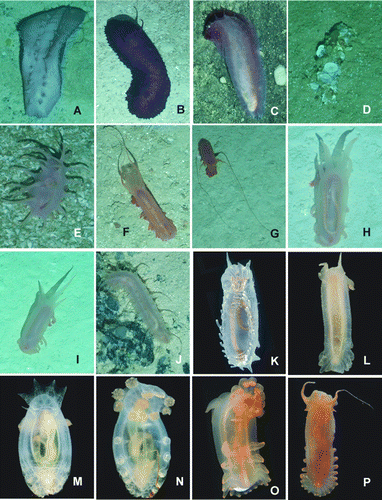
Figure 19. Freshly caught specimens from trawls and ROV, photos courtesy: David Shale. (A) Abyssocucumis abyssorum, St. JC037/15; (B) Benthothuria funebris, St. JC037/15; (C) Amperima furcata, St. JC048/54 Dive 179; (D) Peniagone azorica, St. JC048/24 Dive 165; (E) Peniagone coccinea sp. nov., St. JC048/54 Dive 179; (F) Ellipinion alani sp. nov., St. JC048/24 Dive 165; (G) Laetmogone billetti sp. nov., St. JC048/56, Dive 180; (H,I) Laetmogone billetti, St. JC048/24 Dive 165; (J) Kolga nana, St. JC048/24 Dive 165; (K) Ellipinion delagei, St. JC048/24 Dive 165; (L) Gephyrothuria alcocki, St. JC037/61.
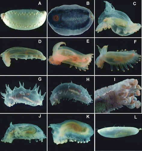
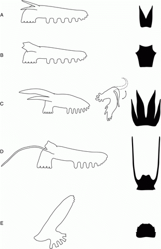
Figure 20. The role of the velum in locomotion. (A) Pressing force; (B) lifting force.
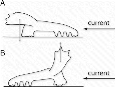
Figure 21. Morphological diversification in Peniagone. Side view (left row) and shape of the velum (right row). (A) Peniagone azorica and Peniagone islandica; (B) Peniagone horrifer; (C) Peniagone coccinea sp. nov.; (D) Peniagone longipapillata; (E) Peniagone diaphana.