
Figures & data
Table I. Morphological characters and states used for identification in Coralliidae.
Table II. List of coralliid species as a result of the taxonomic revision.
Figure 1. Corallium stylasteroides. USNM 77450: (A) Different sides of colony with enlargements of (a) cortex, (b) autozooids and (c) main stem; (B) SEM of cortex; (C) SEM of axis; (D) SEM of cortical mound and retracted autozooid. Black arrows in B indicate siphonozooids.
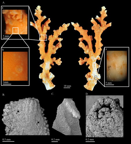
Figure 2. Corallium stylasteroides.USNM 77450: (A) Cortical sclerites from the tip of the colony, (a) 6-radiates, (b) 8-radiates, (c) double clubs; (B) cortical sclerites from the base of the colony, (a) 7-radiates; (C) sclerites from cortical mound and autozooid.
Figure 3. (A) Hemicorallium reginae. Syntype, NBC COEL_02395; (B) Hemicorallium halmaheirense. Syntype, NBC COEL_02393, large fragment, white arrow indicates polychaetes in the middle of the branch; (C) Hemicorallium halmaheirense. Syntype, NBC COEL_02393, small fragment and enlargement of autozooids.
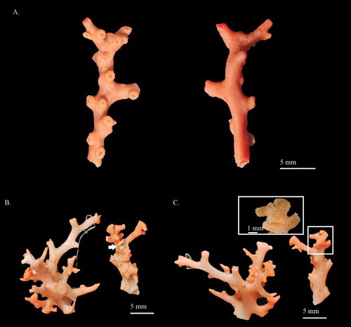
Figure 4. Hemicorallium reginae. Syntype, NBC COEL_02395: (A) Cortical sclerites, (a) crosses, (b) asymmetrical 6-radiates, (c) 7-radiates, (d) symmetrical 8-radiates, (e) double club, (f) irregular forms; (B) sclerites from autozooid, (a) long spindles.
Figure 5. Hemicorallium halmaheirense. Syntype, NBC COEL_02393: (A) Cortical sclerites, (a) spherical 8-radiate; (B) sclerites from autozooid.
Figure 6. Hemicorallium imperiale. Holotype, USNM 50110: (A) Front, back and enlargement of colony; (B) cortical sclerites, (a) elongated 8-radiates reaching 0.1 mm in length.
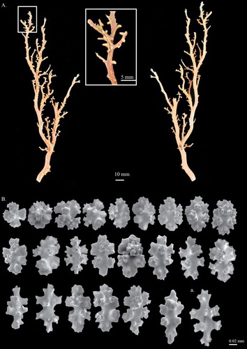
Figure 7. Hemicorallium aurantiacum sp. nov. (A)–(D) Holotype, MNHN-IK-2011-1580: (A) Front of colony; (B) enlargement of frontal colony, white arrows indicate the thickened cortex induced by polychaetes; (C) and (D) SEM of autozooids, white arrow indicates the apical pore of a siphonozooid; (E) paratype, USNM 1196454, front of colony.
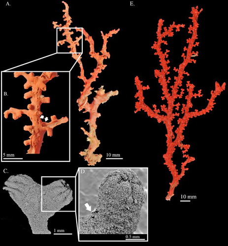
Figure 8. Hemicorallium aurantiacum sp. nov. Holotype, MNHN-IK-2011-1580: (A) Cortical sclerites, (a) 6-radiates, (b) criss-cross, (c) 7-radiates, (d) spherical 8-radiates; (B) sclerites from autozooid, (a) spherical 8-radiates, (b) 7-radiates, (c) spiny spindles, (d) long spindles.
Figure 9. Hemicorallium ducale. Holotype, USNM 50111: (A) Front of colony; (B) SEM image of branch; (C) sclerites from autozooids. Branch image by F.M. Bayer courtesy of Stephen Cairns.
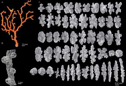
Figure 10. Hemicorallium guttatum sp. nov. Holotype, USNM 1072452 (A)–(C): (A) Front and back of colony; (B) SEM of branch and enlargement of autozooid; (C) SEM of the inner structure of autozooid and enlargement of partial cross-section of tentacle; (D) paratype, USNM 1116818; white arrows indicate the thickened cortex induced by polychaetes.
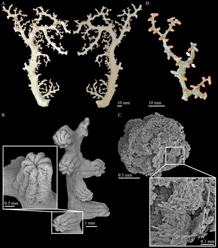
Figure 11. Hemicorallium guttatum sp. nov. Holotype, USNM 1072452: (A) Sclerites from cortex, (a) spherical 8-radiates; (B) sclerites from autozooid, (a) spherical 8-radiates.
Figure 12. (A) Pleurocorallium porcellanum. Holotype, IORAS IV-9-Alc-11-009; (B) Pleurocorallium kishinouyei. Holotype, USNM 94462; (C) Pleurocorallium kishinouyei. Paratype, USNM 94463.
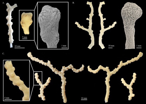
Figure 13. Pleurocorallium porcellanum. Holotype, IORAS IV-9-Alc-11-009: (A) Cortical sclerites; (B) sclerites from autozooid.
Table III. Main characters of Pleurocorallium porcellanum (Pasternak, Citation1981) and Pleurocorallium kishinouyei (Bayer, Citation1996).
Figure 14. Pleurocorallium bonsaiarborum sp. nov. Holotype, MNHN-IK-2009-942: (A) Different sides of colony; (B) cortical sclerites; (C) sclerites from autozooid.
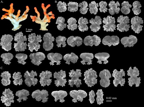
Figure 15. Pleurocorallium clavatum sp. nov. Holotype, USNM 1195223: (A) Front and back sides of colony, and enlargement of branch; (B) SEM image of branch and cross-section.
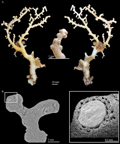
Figure 16. Pleurocorallium clavatum sp. nov. Holotype, USNM 1195223: (A) Cortical sclerites, (a) 6-radiates similar to the radiation warning symbol, (b) double clubs with smooth surface; (B) sclerites from autozooid, (a) rods in pharyngeal region.
Figure 17. Pleurocorallium borneense. Holotype, USNM 49325: (A) Front and back views of colony, and enlargement of branch; (B) sclerites from cortex, (a) double clubs with smooth surface.
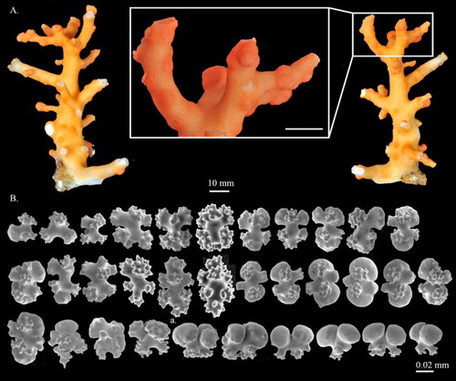
Figure 18. Pleurocorallium norfolkicum sp. nov. Holotype, MNHN-IK-2011-1467 (A)–(C): (A) Front and back sides of colony; (B) close-up of cortical mounds and autozooids, white and black arrows indicate cortical mounds and openings of siphonozooids, respectively; (C) deep autozooid pits with beaded margins; (D) paratype, MNHN-IK-2011-1706.
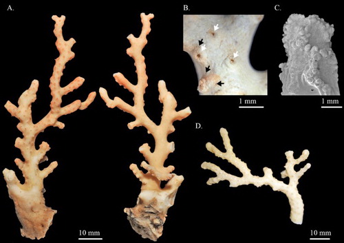
Figure 19. Pleurocorallium norfolkicum sp. nov. Holotype, MNHN-IK-2011-1467: (A) Cortical sclerites from the tip of the colony, (a) symmetrical 6-radiates, (b) asymmetrical 6-radiates, (c) 7-radiate, (d) mushroom-like double clubs, (e) regular double clubs; (B) cortical sclerites from the base of the colony, (a) 7-radiates; (C) sclerites from cortical mounds and autozooid, (a) spherical 6-radiates, (b) small rods in pharyngeal region.
Figure 20. Multiple factor analysis (MFA) scatterplot of 26 Hemicorallium specimens based on both qualitative and quantitative morphological features.