Figures & data
Figure 1. Diagrammatic illustrations (with photographic documentation) of pore filament and canal shape types in Mesophyllaceae: a, non-differentiated pore filaments in straight canals (Athanasiadis and Adey Citation2006, figure 15, Leptophytum tenue, RWM); b, development of cell bars (arrows) from basal cells in Phragmope discrepans (Athanasiadis Citation2020, e); c, thinner-wider pore cells, in transverse (white arrows) or tangential (arrowheads) sections, or in views from the roof interior (black arrows), in Mesophyllum spp. (Athanasiadis Citation2007b, figures 63, 77 Mesophyllum crassiusculum, M. megagastri; RWM); d, thinner-wider pore cells, shorter than adjacent roof cells in Mesophyllum lichenoides (Athanasiadis and Neto Citation2010, figures 25, 29, RWM); e, elongate subbasal cells (shadowed), in certain species of Mesophyllum-Leptophytum (Athanasiadis and Adey Citation2006, figure 47, L. lamellicola, RWM); f, Thallis-type: basally branched pore filaments (supporting a roof filament, black arrows) composed of elongate subbasal cells (shadowed) and two or three smaller cells lacking epithallial cell and terminating below the conceptacle surface (arrowheads) (Keats and Maneveldt 1997, figure 17, as M. incisum, RWM; Figures 4e–h, 9g–i, 12d–e); g, Printziana-type, in P. australis (c–h) and differing from Thallis in having in addition elongate basal cells, and occasionally terminating at the roof surface or just below; h, Sunesonia-type, in S. pseuderubescens (l–p) and differing from Printziana by the development of 4-celled pore filaments terminating below the roof surface, and in having reduced (or deteriorated) basal cells; i, Melyvonnea-type in species of this genus (Jesionek et al. Citation2016, figure 9F, RWM), where pore filaments are unbranched, composed of elongate basal cells (arrows) supporting 2-4 smaller cells and terminating in an epithallial cell below the roof surface; j, triangular (conical) canal with thinner-wider cells along half the canal length (arrows) (Athanasiadis Citation2018b, figure 54, Synarthrophyton patena, RWM); k–m, pyriform canals with thinner-wider basal and subbasal cells (arrows) (k–l: Athanasiadis Citation2018a, figures 30, 31, Carlskottsbergia antarctica, RWM); m, swollen and deeply staining differentiated pore cells (arrows) (Ricker Citation1987, figure 73h, RWM). Abbreviations: canal (c), pore filament (pf), sunken pore filament (spf), reproduced with modifications (RWM); black squares define epithallial cells, and elongate pore cells are shadowed.
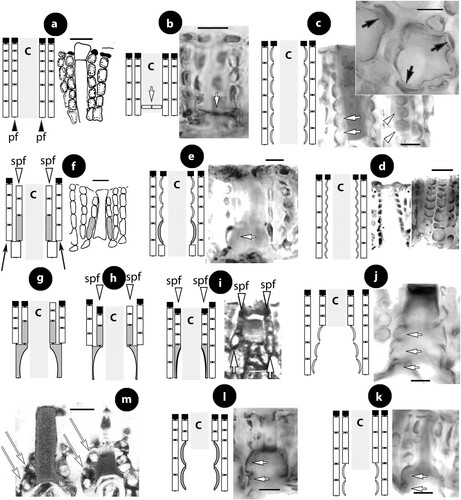
Figure 2. Thallis capensis gen. & sp. nov. Vegetative and spermatangial structures; a–b, tetrasporophyte with flattened multiporate conceptacle roofs (holotype); c–d, carposporophyte with conical uniporate conceptacles (syntype); e, thallus section showing a non-coaxial hypothallium (black arrow) with descending hypothallial (long black arrow) and ascending stratified perithallial filaments (white arrow) (isotype); f, thallus section showing a core of at least two or three cell layers (arrows) running parallel to the substratum and giving rise to ascending perithallial (p) and descending (d) hypothallial filaments (isotype); g, surface section showing elongate subepithallial cells (arrows), just divided (white arrowheads), supporting single epithallial cells (black arrowhead) (isotype); h, descending hypothallial filaments ending in wedge-shaped cells (arrowheads). Note the cell fusion (arrow) (isotype); i, spermatangial structures. SMCs (black arrow) cut off elongate spermatangia (arrowhead) which liberate spermatia (white arrow) (Keats and Maneveldt Citation1997, figure 7, reproduced with modifications). Abbreviations: perithallial filaments (p), descending hypothallial filaments (d).
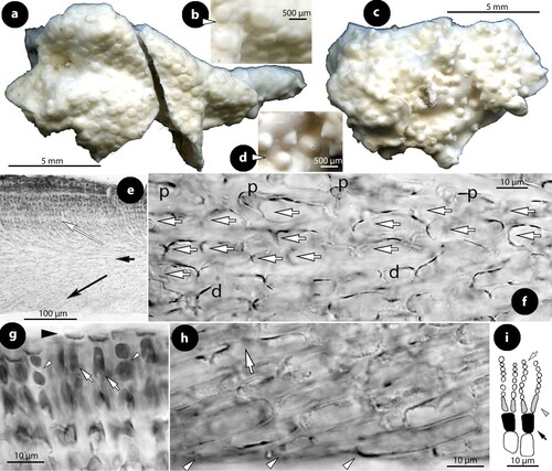
Figure 3. Thallis capensis gen. & sp. nov. Carpogonial, postfertilization stages and carposporangial conceptacle structures (syntype); a, embedded, unfertilized carpogonial conceptacle; b–c, 2- or 3-celled carpogonial branches composed of a carpogonium (ca), a hypogynous (h) and a supporting (s) cell. Note the cell tube (c.t.) connecting the base of the carpogonium with the supporting cell; d–e, postfertilization stages showing the development of a fusion (f) cell that incorporates at least six supporting cells and one hypogynous cell (arrow in figure e), while carpogonia (ca) and basal (b) cells remain intact. Adjacent hypogynous cells develop a contact (broken arrows) to the fusion cell or to gonimoblast filaments (g.f.); f–g, carposporangial conceptacles with a distinctively or slightly raised fertile zone (arrows) and peripheral production of carposporangia (arrowheads); h, older carposporangial conceptacles embedded in the perithallium. Note the lack of a raised fertile zone. Abbreviations: b (basal cell), ca (carpogonium), c.t. (cell tube), f (fusion cell), g.f. (gonimoblast filaments), h (hypogynous cell), s (supporting cell).
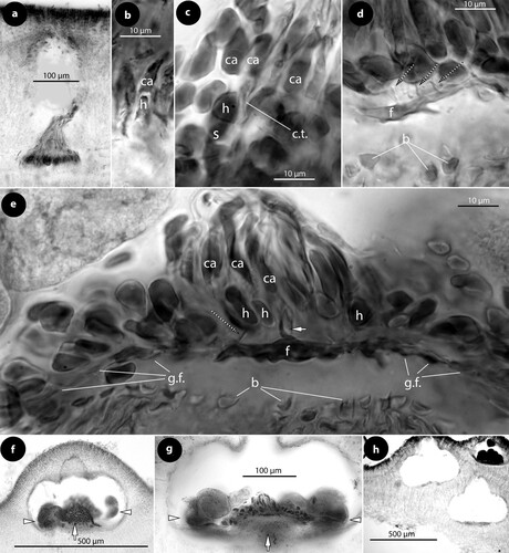
Figure 4. Thallis capensis gen. & sp. nov. Tetrasporangial conceptacle structures (isotype); a, tetrasporangial conceptacle with three tetrasporangia; b–d, views of a multiporate roof from above showing pore (p) canals surrounded by 6–7 pore cells; e–h, sections of canals of multiporate roofs showing filaments lining the canals. Note the branched basal cells (long white arrows), each supporting a normal roof filament (black arrows) and the rest of the lining filament that is composed of an elongate subbasal cell (short white arrows) followed by two cells (arrowheads). Pore filaments lack epithallial cells and terminate below the roof surface, i.e. epithallial cells (black arrowheads) of adjacent roof filaments; i, a diminutive, 3-celled, filament (broken arrow) at the canal base. Abbreviations: p (pore canal).
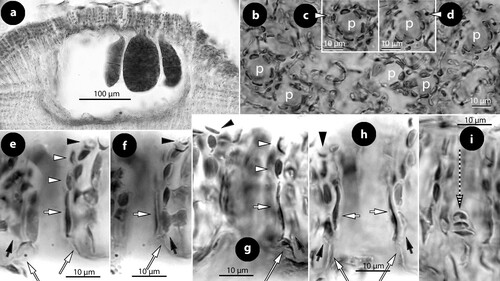
Table I. Comparative data between Thallis, Perithallis, Printziana, Sunesonia and related genera and species of Mesophyllaceae.
Figure 5. Perithallis incisa gen. & comb. nov. Original material in TRH (B17-2551, B17-2552, B17-2553); a, the lectotype collection (B17-2551) that includes materials in a box (A) holding a Setchell label (B), remains of the collection in small fragments (C), two Foslie slides (n°1175; D, E), and a smaller box (F) that includes the here selected lectotype specimen (G); b, the here selected lectotype, a conglomerate including two thalli with multiporate conceptacles (top) and gametangial thalli underneath, mostly carposporophytes (arrowhead); c, thalli with multiporate and a few uniporate (male; arrowhead) conceptacles (syntype B17-2552); d, thallus belonging to a different species provided with smaller carpogonial-carposporangial conceptacles (syntype B17-2553); e, side view of a broken unattached thallus of the lectotype showing coaxial hypothallial growth; f, view of the underside of a carposporangial thallus, showing zonate texture (lectotype); g, view of the underside of a male thallus, showing smooth texture (lectotype).
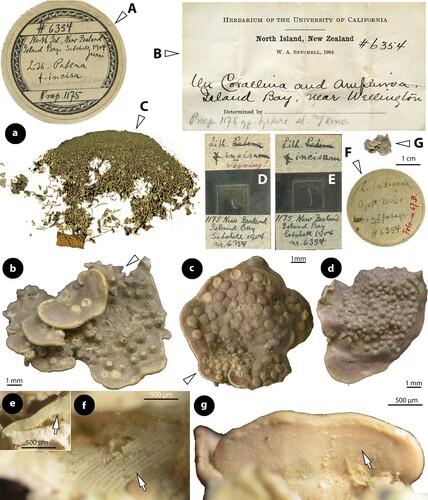
Figure 6. Perithallis incisa gen. & comb. nov. Vegetative structures; a, section at the margin showing a non-coaxial patch (black arrow) between two coaxial ones (white arrows) (iso-lectotype); b, section of a carposporophyte with predominantly coaxial growth (iso-lectotype); c, surface view of a decalcified lamella showing a striated thallus (due to coaxial hypothallial growth) (syntype, Setchell n°6354 in TRH, B17-2551); d, thallus section showing a core of up to four cell layers of hypothallial filaments running parallel to the substratum, supporting ascending perithallial (p) and descending (d) hypothallial filaments (LTB 14763). Abbreviations: p (perithallial filaments), d (descending hypothallial filaments).
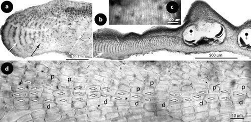
Figure 7. Perithallis incisa gen. & comb. nov. Vegetative structures; a, terminal meristematic cells (arrows) protected by a cuticle (c) undergoing synchronous anticlinal divisions (LTB 14763); b, section at the margin showing terminal meristematic cells (white arrows) progressively displaced dorsally to become epithallial cells (arrowheads), at the end of the protective cuticle (c). Note the subdichotomous hypothallial divisions (black arrows) that cause the thallus vertical expansion (syntype B17-2551); c, section at the dorsal surface showing elongate subepithallial cells (white arrows) and a layer of flattened epithallial cells (arrowheads) (LTB 14763); d, terminal meristematic cells (arrow) progressively displaced ventrally to become epithallial cells (arrowheads) (syntype, B17-2551); e, section at the ventral side showing descending (d) hypothallial filaments ending in wedge-shaped cells (arrows) (iso-lectotype); f, section at the ventral side of a tetrasporangial specimen, showing descending filaments (d) terminating into perithallial cells (p) supporting single flattened epithallial cells (arrowheads). Note the trichocyte (tr) and the distinctive angle between perithallial (p) and descending (d) hypothallial cells (LTB 14763); g, section at the ventral side showing descending filaments (d) to support one layer of perithallial cells (arrows) with terminal epithallial cells (arrowheads) (iso-lectotype); h–i, sections at the surface showing flattened epithallial cells (black arrowheads), an elongate subepithallial cell (arrow), a trichocyte (tr), and the presence of two epithallial cells in sequence (white arrowheads) (LTB 14763). Abreviations: c (cuticle), d (descending hypothallial filament), p (perithallial cell), tr (trichocyte).
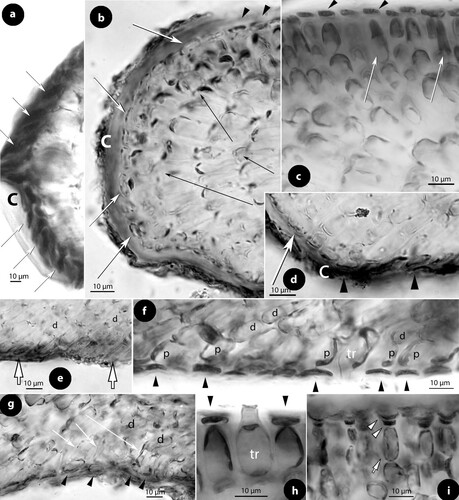
Figure 8. Perithallis incisa gen. & comb. nov. Male, carpogonial and postfertilization structures (LTB 14763); a, section at the chamber floor of a mature male conceptacle, showing lunate SMCs (arrows) cutting off spermatangia (arrowheads) that release roundish spermatia; b, section at the chamber floor of an old male conceptacle, showing remains of SMCs (arrows) supporting elongate or inflated, empty spermatangia (arrowheads); c–d, carposporangial conceptacles showing peripheral development of carpospores (arrows) from a centrally raised (black arrowhead) or nearly flattened (white arrowhead) fertile zone; e, section at the carposporangial floor showing peripheral production of carposporangia (c) from a gonimoblast filament (black arrow), and the presence of gonimoblast filaments (white arrows). Note the remains of carpogonia (ca) and hypogynous cells (h), which remain intact in contrast to basal (b) cells that are incorporated in the gonimoblast filaments; f–g, section at the carposporangial floor showing remains of carpogonia (ca) and hypogynous cells (h) that remain intact. Gonimoblast filaments (long white arrows) form a chain of smaller fusions cells, each including one or two supporting cells and their basal cells. Carposporangia (c) are produced from the periphery (black arrow). Gonimoblast filaments bend down (black arrow) from an apparently raised fertile zone. The connection (normal pit-plug?) between a hypogynous cell and the gonimoblast filament (broken line) is magnified in figure g; h, view of the carposporangial floor from above, at the level of carposporangia (c), showing their peripheral production and the apparent lack of a fusion cell. Abbreviations: b (basal cell), c (carposporangium), ca (carpogonium), h (hypogynous cell).
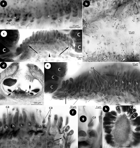
Figure 9. Perithallis incisa gen. & comb. nov. Multiporate conceptacle structures; a–b, sections showing two thalli (arrows) growing back-to-back, one provided with a multiporate conceptacle. The presence of ventral epithallial cells in the upper thallus is magnified in figure b (arrowhead) (Foslie slide n°1175 ‘Tegning’, syntype B17-2551); c, two tetrasporangia (LTB 14763); d–e, surface views of pore canals of a multiporate roof, surrounded by 11 respectively nine rosette (r) cells (syntype B17-2551); f–i, sections of canals of multiporate roofs, showing pore filaments lining the canals. Basal cells (white long arrows) are branched supporting a normal roof filament (long black arrows) and the rest of the lining filament that includes an elongate subbasal cell (white arrows) and two or three top cells (white arrowheads) lacking epithallial cells (that occur in adjacent roof filaments; black arrowheads) and terminating below the roof surface (f: Woelkerling and Harvey Citation1993, figure 14D ‘Lectotype collection’, reproduced with modifications; g: syntype B17-2551; h–i: LTB 14763). Abbreviations: r (rosette cell).
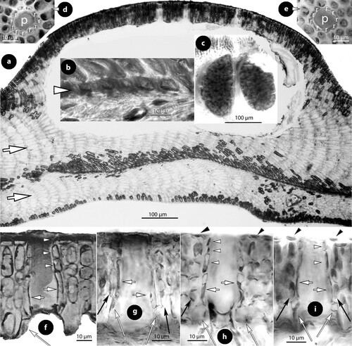
Figure 10. Perithallis chathamensis (Foslie) comb. nov. The original material in TRH (B18-2594) that comprises materials placed in a box (a) and including: four paper sheets (b, c, d, e), three slides (f, g, h), two smaller boxes (i, j) with minute algal fragments, and a single large specimen (a), the here selected lectotype (scale bars: a–j: 2 cm; k: 1 cm). Two magnifications of the lectotype (l, m) show multiporate conceptacles (arrows).
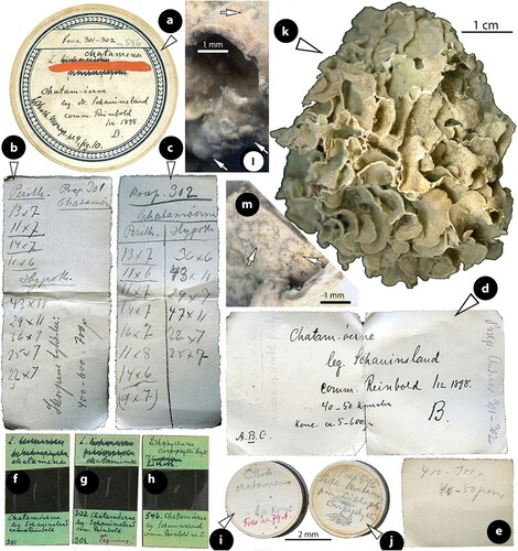
Figure 11. Perithallis chathamensis (Foslie) comb. nov. Vegetative structures; a, section showing a predominantly coaxial hypothallium (short white arrow) supporting ascending perithallial filaments (black arrow) and descending hypothallial filaments (long white arrow). Note the multiporate conceptacle with slightly sunken roof (arrowhead) (NZC0745); b, view of a decalcified thallus from above showing the predominantly coaxial hypothallium (NZC0745); c, section at the margin showing terminal synchronous, anticlinal divisions (black arrows) below the cuticle (white arrows) (NZC0745); d, section at the surface where terminal meristematic cells (arrows) become epithallial cells (arrowheads). The cuticle (white arrows) ceases to exist (protect) the meristematic cells at the transition zone (NZC0745); e, section showing coaxial hypothallial growth (arrow) (iso-lectotype); f, section of two lamellae (arrows) growing back-to-back (iso-lectotype); g, conglomerate of several lamellae (arrows) anastomosing. Not clear if all lamellae belong to the same individual/species (Foslie slide n°302, B18-2594); h, section showing two thalli growing back-to-back (arrows) and gradually becoming confluent (NZC0745); i–j, sections at the ventral side showing isodiametric to flattened epithallial cells (arrowheads) terminating 1- or 2-celled perithallial (p) filaments. Note the trichocytes (tr) and the angle between descending hypothallial (h) and perithallial (p) cells (i: iso-lectotype; j: NZC0745); k–m, sections at the surface showing epithallial cells (arrowheads) and trichocytes (tr). Note that subepithallial cells are longer than cells below (k: iso-lectotype; l–m: NZC0745).
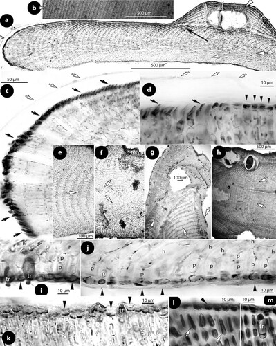
Figure 12. Perithallis chathamensis (Foslie) comb. nov. Multiporate conceptacle structures; a–b, sections of multiporate conceptacles showing the roof with one canal (arrowhead) and a second roof (Figure b) with three canals (c) (iso-lectotype); c, surface view of a canal showing nine rosette (r) cells surrounding the pore (p) (iso-lectotype; Keats and Chamberlain Citation1997, figure 10, redrawn with modifications); d–e, sections of two canals showing 4-celled lining filaments, with branched basal cells (long white arrows) supporting a roof filament (black arrows), and the rest of the lining filament that is composed of elongate subbasal cells (white arrows), and two more cells (arrowheads) that lack epithallial cells ending below the conceptacle surface. Note that adjacent roof filaments are provided with epithallial cells (black arrowheads) (d: NZC0745; e: iso-lectotype). Abbreviations: c (canal), r (rosette cell).
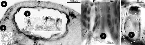
Figure 13. Printziana australis gen. & nom.nov. Vegetative structures; a, thalli in view from above and the side (arrows) showing the prominent erect perithallial protuberances (isotype); b, transverse section of a thallus encircling an object (arrowhead) and producing a perithallial protuberance (arrow). Note the embedded tetrasporangial conceptacles (isotype); c, transverse section of a perithallial protuberance with embedded tetrasporangial conceptacles (LTB 12850); d, section at the margin showing synchronous anticlinal divisions in terminal hypothallial cells (white arrows) protected by a cuticle (c). Subdichotomous divisions (black arrow) expand the thallus and terminal meristematic cells become displaced developing to epithallial cells (arrowheads) – lower part magnified in figure f (NZC2508); e, section showing a predominantly coaxial, arching hypothallium (arrow) supporting an ascending stratified (black arrow) perithallium (isotype); f, section near the ventral side showing a subdichotomous division (black arrow) and descending hypothallial filaments ending in wedge-shaped cells (arrowheads) (NZC2508); g–j, sections at the surface showing elongate (long white arrows) or just divided (white arrows) subepithallial cells, supporting single epithallial cells; the latter spherical to flattened or even elongate (black arrowheads). Note two terminal trichocytes (black arrows) and the fusions between contiguous (arrowheads) cells (g–h: isotype; i: LTB 12850; j: NZC0621). Abbreviations: c (cuticle).
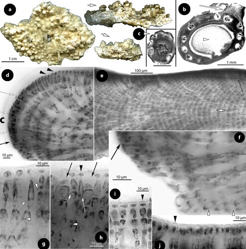
Figure 14. Printziana australis gen.& nom. nov. Male structures (isotypes); a–b, early stages of simple (unbranched) spermatangial structures restricted to the floor with lunate SMCs (arrows) (Woelkerling and Harvey Citation1993, figure 29B, reproduced with modifications); c–d, male conceptacle at late maturity with elongate SMCs (broken arrows) supporting spermatangia in pairs (arrowheads) that release spermatia (black arrowheads).
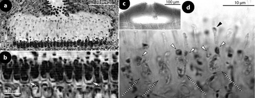
Figure 15. Printziana australis gen.& nom. nov. Carpogonial, postfertilization and carposporangial structures (NZC0621); a–b, carpogonial conceptacle with unfertilized carpogonial branches extending across the entire floor. Carpogonial branches composed of carpogonia (ca) and hypogynous (h) cells, attached to supporting (s) cells. Note that supporting cells support the chamber walls (arrows) when they do not support carpogonial branches; c, a carpogonial branch composed of a carpogonium (ca) and a hypogynous (h) cell, attached to a supporting (s) cell. A sterile cell may occur attached to the hypogynous cell (broken line); d, postfertilization stage showing a distinctive fusion cell (f), composed of at least 6 or 7 supporting cells and incorporating at least 2 hypogynous (h) cells, producing the gonimoblast filaments (g.f.). Remains of carpogonia (ca), hypogynous (h) and basal (b) cells remain intact and do not merge with the fusion cell or the gonimoblast filaments; e–f, peripheral production (arrows) of carposporangia (c) from a slightly or distinctively raised fertile zone (pe). Abbreviations: b (basal cell), c (carposporangia), ca (carpogonium), g.f. (gonimoblast filament), h (hypogynous cell), pe (pedestal), s (supporting cell).
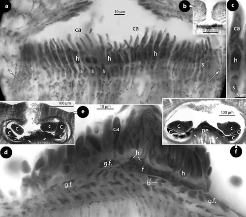
Figure 16. Printziana australis gen.& nom. nov. Tetrasporangial structures (a–h) and Printziana spp. (i–k); a, section of a tetrasporangial conceptacle with sunken roof (isotype); b, surface view of canals (p) of a multiporate roof surrounded by seven-nine rosette cells (NZC0621); c–h, sections through canals of multiporate roofs showing elongate basal (long white arrows) and subbasal (short white arrows) cells, each supporting two (occasionally three) cells (arrowheads) the last being occasionally an epithallial cell (black arrowheads). Note that the branched basal cells also support a normal roof filament (black arrows) (c–e: isotype: f–h: NZC0621); i, bisporangial thallus with 5-celled pore filaments composed of elongate basal and subbasal cells (arrows) and three smaller cells (arrowheads) (NZC0858); j, tetrasporangial thallus with at least 6-celled pore filaments composed of elongate basal and subbasal cells (arrows) and four smaller cells (arrowheads) (NZC2141); k, bisporangial thallus with ‘multi-rim’ outgrowths (arrowheads) around the sunken pore plate (NZC2269). Abbreviations: p (pore canal).
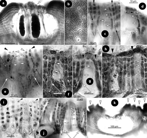
Figure 17. Sunesonia pseuderubescens, gen. & sp. nov. Vegetative and multiporate conceptacle structures; a, habit of the holotype from above showing several erect perithallial protuberances (arrows); b, section at the margin showing hypothallial meristematic cells (black arrows) producing a non-coaxial hypothallium (white arrow) (isotype); c, section showing descending hypothallial filaments ending in wedge-shaped cells (arrowhead)(isotype); d, section at the surface showing ± stratified perithallium (isotype); e–f, sections at the thallus surface showing isodiametric to flattened epithallial cells (arrowheads), supported by elongate (arrows) subepithallial cells (isotype); g, section of a tetrasporangial conceptacle (NZC0061); h–i, transverse sections through an erect perithallial protuberance showing superficial and embedded (arrows) conceptacles with bisporangial remains (isotype; scale bar: 500 µm); j, an empty conceptacle with distinctive convex roof (isotype); k, surface view of a multiporate roof showing canals (p) surrounded by 7–9 rosette cells (LTB 14744); l–p, sections through canals of multiporate roofs showing 4-celled lining filaments composed of elongate basal (long white arrows) and subbasal (short white arrows) cells, followed by two smaller cells (white arrowheads). Top cells can be epithallial cells (double arrowhead) and generally end below epithallial cells (black arrowheads) of adjacent roof filaments. Basal cells are divided supporting a roof filament (black arrows) and the rest of the pore filament. Note that basal cells are ± reduced. Figures o–p are tangential sections (facing pore filaments) (l–m, p: isotype; n–o: NZC0835). Abbreviations: p (pore canal).
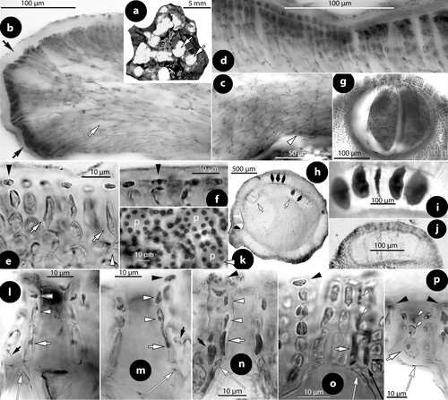
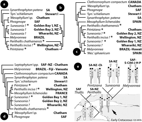
Figure 18. a–d, relationships between species and genera of Mesophyllaceae as indicated by nSSU (a, c, d) and psbA (b) gene sequences, using maximum likelihood (a, d) and Bayesian inference (b, c); a–b, after Broom et al. (Citation2008, table 2, figure 5, figure 1); c, after Peña et al. (Citation2011, figure 3); d, after Bittner et al. (Citation2011, table 2, figure 1). Unidentified taxa in the cited papers have been excluded in the here shown reproductions. Ingroup members polarized by the closest outgroup taxon: Synarthrophyton spp. (a, b), Clathromorphum compactum (c), and ‘Leptophytum’ spp. (d). Phragmope replaces a sequence of ‘Mesophyllum engelhartii’ from South Africa (Athanasiadis Citation2020, figure 8); Melyvonnea replaces sequences of Mesophyllum erubescens from Brazil, Hawaii, Fiji and Vanuatu (Athanasiadis & Ballantine Citation2014, figure 2D). New taxa proposed in this study are indicated by asterisks and replace sequences of Mesophyllum printzianum (= Printziana) or ‘Mesophyllum erubescens’ (= Perithallis chathamensis, P. incisa ?, Sunesonia ?) from New Zealand-Chatham. Accession numbers (in alphabetic order): Clathromorphum compactum Canada (U60742); ‘Leptophytum’ spp. SAF (U62119 & U62120); ‘Leptophytum’ repandum NZ (EF628216); ‘Leptophytum’ repandum Chatham (EF628215); Melyvonnea Brazil (U61257); Melyvonnea Hawaii (DQ629011, DQ629012); Melyvonnea Fiji-Vanuatu (GQ917390, GQ917392, GQ917411); Mesophyllum lichenoides Spain, France (CH13992, GQ917384); ‘Mesophyllum’ sphaericum Spain (CH1814, CH1891); ‘Mesophyllum’ sp. Chatham (EF628218); Perithallis chathamensis Chatham (EF628222); Perithallis incisa ? Wellington (EF628223); Phragmope SAF (U61256); Printziana Chatham (EF628224); Sunesonia ? Wharariki Beach, Golden Bay 1, 2 (EF628219, EF628220, EF628221); Synarthrophyton patena SA (U61255); ‘Synarthrophyton’ schielianum Stewart Island (EF628217). e, hypothetical evolution of pore filaments in Thallis, Perithallis, Printziana, Sunesonia and Melyvonnea, based on parsimony. The ancestral condition, Thallis-type (white arrow), persisted in Thallis-Perithallis. A derivative condition (black arrow), involving the acquisition of elongate basal cells, is the ancestor of the present-day Printziana-, Sunesonia- and Melyvonnea-types. It assumes that the Melyvonnea-type evolved via loss of basally branched pore cells, the latter persisted in Printziana and reduced in Sunesonia. Black squares indicate epithallial cells and arrowheads sunken pore filaments (below the conceptacle roof). Abbreviations: c (canal), SA (southern Australia), NZ (New Zealand), Ch (Chatham), SAF (South Africa), S.-C.Atl. (South & Central Atlantic), J (Japan), H (Hawaii), P (Polynesia).