Figures & data
Figure 1. Schematic drawing of Mesodinium rubrum at rest (top) and during a jump (bottom). The arrows at the bottom of the jumping cell indicate the direction of jumping and the direction of cell rotation. The arrows along the side indicate the direction of the effective stroke of the membranelles.
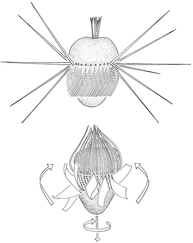
Figure 2. The position of cirri and membranelles during a jump traced from a high-speed video camera recording. The numbers indicate milliseconds after initiation of the jump.
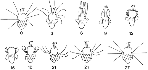
Figure 3. Velocity and distance travelled during a jump.
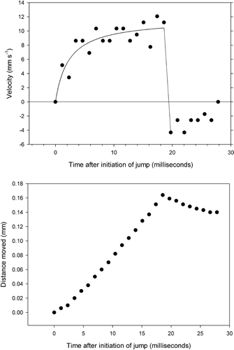
Figure 4. The behaviour of Mesodinium cells in a siphon flow. The numbers refer to fluid velocity in mm s−1. Left: tracks of two ciliates in the flow. The dotted lines are the passive movement along flow lines; the solid lines are jumps. Right: initiation of jumps (solid circles) and of sustained swimming against the flow (triangles). The internal opening of the capillary is 0.15 mm.
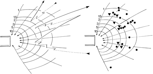
Figure 5. Angles of successive jumps of five Mesodinium cells relative to flow lines in a siphon flow. A value of 0° means swimming directly along the flow and a value of 180° that the cells swim directly against the current.
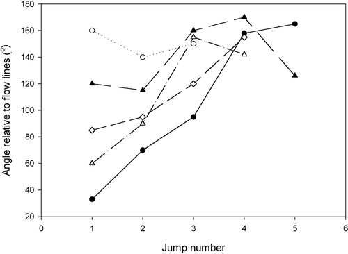
Figure 6. The behaviour of a cell in a siphon flow traced from a high-speed video recording. The cell does not reorientate while flowing passively along flow lines (solid lines), but reorientates against the flow at the initiation of jumps by activating the cirri in the leading edge of the cell. The time interval between dots is 1.5 ms.
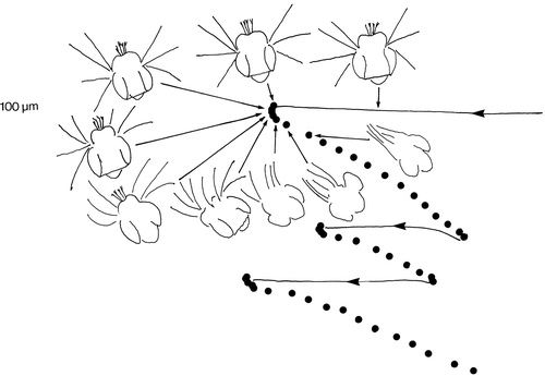
Figure 7. Flow velocity and shear in the siphon flow shown in as a function of distance from the tip of the capillary. The horizontal bars in the right frame show the range of shear that induces the first jumps, and the range of shear that induces sustained swimming against the water flow, respectively.
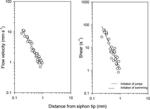
Figure 8. Orientation of the jumping direction relative to the vertical in cells drifting upwards and of cells drifting downwards (right). The reason why the latter drifted downwards is the low frequency of jumping combined with gravitational sinking.
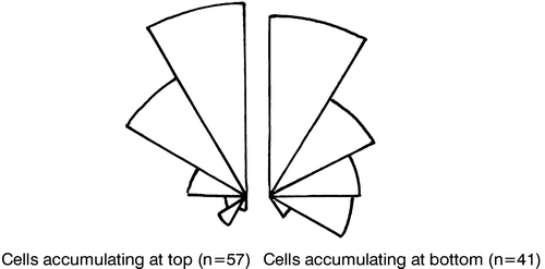
Figure 9. Jumps (solid lines) and sinking (dotted lines) for a cell with a high jumping frequency (left) and for a cell with a low jumping frequency (right).
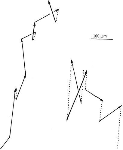
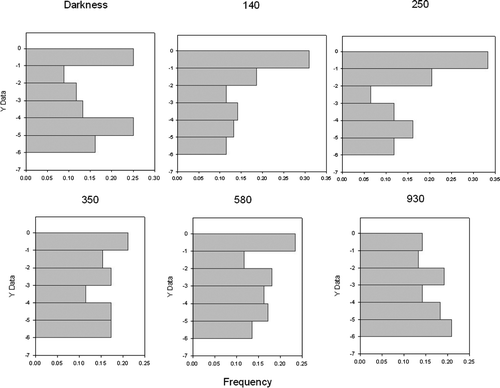
Figure 10. Vertical distribution of Mesodinium in a 6 mm high water column at different irradiance levels (numbers above the graphs are µmol photons m−2 s−1 after about 15 min exposure). Each distribution is based on the position of about 100 cells.