788
Views
15
CrossRef citations to date
0
Altmetric
ORIGINAL ARTICLES

Larval development of Mooreonuphis stigmatis (Treadwell, 1922) (Polychaeta: Onuphidae) from the north-east Pacific

&
Pages 6-24 | Published online: 16 Sep 2009

Figures & data

Table I. Summary of different developmental patterns in onuphids (pattern types sensu Paxton (Citation1993) with modifications: I – brooding in the parental tube with direct development; II – viviparity; III – brooding in masses or sacs attached to parental tube; IV – free-spawning with planktonic larvae).

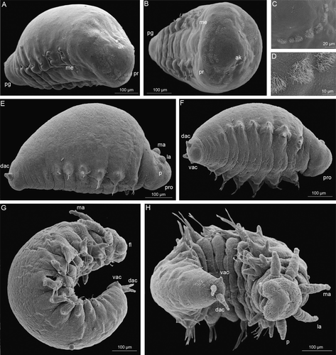
Figure 1.  Early development of Mooreonuphis stigmatis. (A–D) 5-chaetiger larva: (A) lateral view; (B) frontal view; (C,D) fragment of akrotroch. (E,F) 7-chaetiger larva: (E) lateral view, (F) ventral view. (G,H) 11-chaetiger larva: (G) lateral view; (H) ventral view. ak, akrotroch; dac, dorsal anal cirrus; fl, frontal lip; la, lateral antenna; ma, median antenna; me, metatroch; p, palp; pg, pigidium; pr, prototroch; pro, prostomium; vac, ventral anal cirrus.

Figure 1.  Early development of Mooreonuphis stigmatis. (A–D) 5-chaetiger larva: (A) lateral view; (B) frontal view; (C,D) fragment of akrotroch. (E,F) 7-chaetiger larva: (E) lateral view, (F) ventral view. (G,H) 11-chaetiger larva: (G) lateral view; (H) ventral view. ak, akrotroch; dac, dorsal anal cirrus; fl, frontal lip; la, lateral antenna; ma, median antenna; me, metatroch; p, palp; pg, pigidium; pr, prototroch; pro, prostomium; vac, ventral anal cirrus.

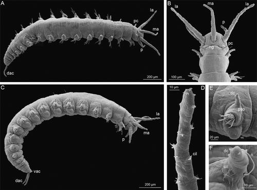
Figure 2.  16-chaetiger larva of Mooreonuphis stigmatis: (A) dorsal view; (B) prostomium, dorsal view; (C) lateral view; (D) median antenna; (E) parapodium of the third chaetiger; (F) dorsal cirrus of the third parapodium. cil, ciliae; dac, dorsal anal cirrus; dc, dorsal cirrus; la, lateral antenna; ma, median antenna; n, notochaeta, ng, nuchal groove, p, palp; pc, peristomial cirrus; pso, parapodial sensory organ; vac, ventral anal cirrus; vc, ventral cirrus.

Figure 2.  16-chaetiger larva of Mooreonuphis stigmatis: (A) dorsal view; (B) prostomium, dorsal view; (C) lateral view; (D) median antenna; (E) parapodium of the third chaetiger; (F) dorsal cirrus of the third parapodium. cil, ciliae; dac, dorsal anal cirrus; dc, dorsal cirrus; la, lateral antenna; ma, median antenna; n, notochaeta, ng, nuchal groove, p, palp; pc, peristomial cirrus; pso, parapodial sensory organ; vac, ventral anal cirrus; vc, ventral cirrus.

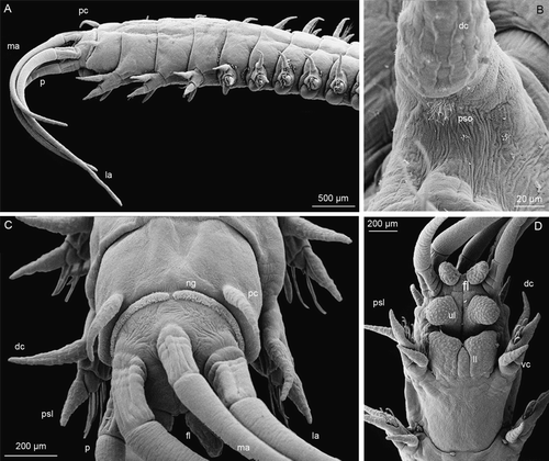
Figure 3.  118-chaetiger juvenile of Mooreonuphis stigmatis: (A) lateral view; (B) parapodial sensory organ of the fifth parapodium; (C) anterior end, dorsal view; (D) anterior end, ventral view. dc, dorsal cirus; fl, frontal lip; la, lateral antenna; ll, lower lip; ma, median antenna; ng, nuchal groove; p, palp; pc, peristomial cirrus; psl, postchaetal lobe; pso, parapodial sensory organ; ul, upper lip; vc, ventral cirrus.

Figure 3.  118-chaetiger juvenile of Mooreonuphis stigmatis: (A) lateral view; (B) parapodial sensory organ of the fifth parapodium; (C) anterior end, dorsal view; (D) anterior end, ventral view. dc, dorsal cirus; fl, frontal lip; la, lateral antenna; ll, lower lip; ma, median antenna; ng, nuchal groove; p, palp; pc, peristomial cirrus; psl, postchaetal lobe; pso, parapodial sensory organ; ul, upper lip; vc, ventral cirrus.

Table II. Different types of provisional larval (lowercase) and permanent adult (uppercase) chaetae in comparison with terminology proposed by other authors.

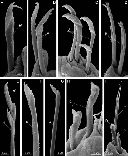
Figure 4.  Chaetae found on different larvae of Mooreonuphis stigmatis: (A) anterior early compound bidentate hooded falciger – b 1 ; (B) anterior pseudocompound bidentate falciger – a; (C) anterior pseudocompound bidentate hooded falciger – b 2 ; (D) anterior pseudocompound tridentate hooded falciger – B; (E) anterior compound bidentate hooded falciger – b 3 and transitional compound bidentate falciger – c; (F,G) transitional compound bidentate falciger – c; (H) posterior compound bidentate subacicular hooded hook – d; (I) posterior compound bidentate subacicular hooded hook – d, simple limbate chaeta – C and posterior simple subacicular bidentate hooded hook – D. Lowercase letters represent provisional larval chaetae, uppercase letters represent permanent adult chaetae.

Figure 4.  Chaetae found on different larvae of Mooreonuphis stigmatis: (A) anterior early compound bidentate hooded falciger – b 1 ; (B) anterior pseudocompound bidentate falciger – a; (C) anterior pseudocompound bidentate hooded falciger – b 2 ; (D) anterior pseudocompound tridentate hooded falciger – B; (E) anterior compound bidentate hooded falciger – b 3 and transitional compound bidentate falciger – c; (F,G) transitional compound bidentate falciger – c; (H) posterior compound bidentate subacicular hooded hook – d; (I) posterior compound bidentate subacicular hooded hook – d, simple limbate chaeta – C and posterior simple subacicular bidentate hooded hook – D. Lowercase letters represent provisional larval chaetae, uppercase letters represent permanent adult chaetae.

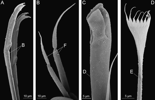
Figure 5.  Chaetae present on adults of Mooreonuphis stigmatis: (A) pseudocompound tridentate hooded falciger – B; (B) compound spiniger – F; (C) large median tridentate hooded hook – D; (D) pectinate chaeta – E.

Figure 5.  Chaetae present on adults of Mooreonuphis stigmatis: (A) pseudocompound tridentate hooded falciger – B; (B) compound spiniger – F; (C) large median tridentate hooded hook – D; (D) pectinate chaeta – E.

Table III. The distribution of different chaetae during the development of Mooreonuphis stigmatis. Lowercase letters represent provisional larval chaetae (a – anterior pseudocompound bidentate falciger, b 1 – anterior early compound bidentate hooded falciger, b 2 – anterior pseudocompound bidentate hooded falciger, b 3 – anterior compound bidentate hooded falciger, c – transitional compound bidentate falciger, d – posterior compound bidentate subacicular hooded hook, n – notopodial capillary chaeta). Uppercase letters represent permanent adult chaetae (A – acicula, B – anterior pseudocompound tridentate hooded falciger, C – simple limbate chaeta, D – posterior simple subacicular bidentate hooded hook, E – pectinate chaeta). Numbers represent the number of each chaeta on the parapodium of corresponding chaetiger.

Table IV. A comparison of the subacicular hooks (SAH) development among ten species of onuphids.

Table V. A comparison of the development of branchiae among five species of onuphids.
Reprints and Corporate Permissions

Please note: Selecting permissions does not provide access to the full text of the article, please see our help page How do I view content?

To request a reprint or corporate permissions for this article, please click on the relevant link below:

Academic Permissions

Please note: Selecting permissions does not provide access to the full text of the article, please see our help page How do I view content?

Obtain permissions instantly via Rightslink by clicking on the button below:

If you are unable to obtain permissions via Rightslink, please complete and submit this Permissions form. For more information, please visit our Permissions help page.