Figures & data
Figure 1. Using the ‘TrimLasso’ brush to select and delete areas of geometry on a micro-CT scan of a skull.
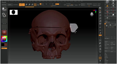
Figure 2. Filling holes with a primitive shape, the ‘Move’ brush can be used to tug the sphere until it fits snuggly in the hole.
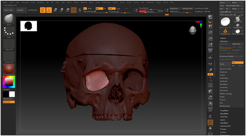
Figure 3. A micro-CT scan of a skull before and after Dynameshing, with the ‘Draw Polyframe’ view switched on.
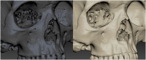
Figure 4. Found in the left shelf, there is a wide variety of Alphas to choose from. These can be useful for painting texture details.
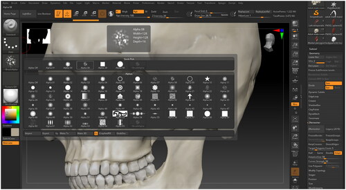
Figure 5. Setting up a texture to paint with using the Spotlight feature.
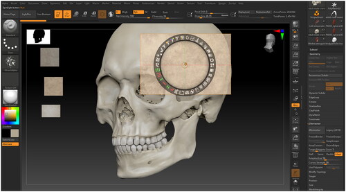
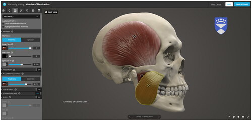
Figure 6. The muscles of mastication have been modelled over this cleaned up micro-CT skull scan.
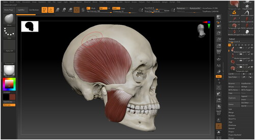
Figure 7. Editing material properties for individual structures in Sketchfab.